Preparatory Mindset
Early pregnancy complications are the most common emergencies in first-trimester obstetrics and a guaranteed exam/OSCE topic. The unifying mindset: any reproductive-age woman with vaginal bleeding ± abdominal pain and a positive pregnancy test has an ectopic pregnancy until proven otherwise. The differential is manageable if you sort by ultrasound and hCG dynamics: ectopic (empty uterus + adnexal mass/pain + abnormal hCG), spontaneous abortion (intrauterine sac ± heartbeat, bleeding, cervical os open/closed), molar pregnancy (GTD) (very high hCG, "snowstorm" scan, no fetus). Management logic: ectopic → methotrexate or surgery; miscarriage → expectant/medical/surgical per stage; molar → surgical evacuation + hCG surveillance (curettage, NOT just observation — molar is premalignant). This chapter also introduces gestational trophoblastic disease (GTD) — the one "cancer" that is almost always curable if you follow hCG to zero.
Core Concepts
1. Differential diagnosis of bleeding in early pregnancy
| Condition | hCG | Ultrasound | Key feature |
|---|---|---|---|
| Threatened abortion | Appropriate/doubling | IUP sac + heartbeat ± subchorionic bleed | Bleeding, closed os, pregnancy viable |
| Inevitable/incomplete abortion | Falling | IUP sac, open os, retained tissue | Progressive bleeding, open os |
| Missed abortion (blighted ovum) | Falling/plateau | Gestational sac without heartbeat / empty sac | No symptoms until late; "anembryonic" |
| Ectopic pregnancy | Slow-rising (<66% in 48 h), plateau, or falling | Empty uterus + adnexal mass/fluid | Pain dominant (unilateral, shoulder-tip, collapse = rupture) |
| Molar pregnancy (complete/partial) | Very high (>100,000 often) | "Snowstorm"/vesicular pattern, no fetus (complete) | Grape-like vesicles; may have preeclampsia <20 wk, hyperemesis |
2. Spontaneous abortion (miscarriage)
- Threatened: rest, reassurance, repeat scan; progesterone in selected cases (recurrent loss, bleeding + previous loss). - Missed/incomplete/inevitable: expectant (up to 14 days), medical (misoprostol ± mifepristone), or surgical (MVA/ERPC) — surgical for haemorrhage, infection, patient choice, or incomplete after medical. - Recurrent loss: investigate — karyotype (both partners), uterine cavity (HSG/USS), antiphospholipid antibodies, thyroid, glucose.
- Definition: pregnancy loss <20 weeks (China: <28 weeks or fetal weight <1000 g).
- Incidence: ~15-20% of clinically recognised pregnancies; ~80% of losses in first trimester; most sporadic losses are chromosomal abnormalities (50-60%).
- Classification: threatened / inevitable / incomplete / complete / missed / recurrent (≥2-3 consecutive losses).
- Risk factors: advanced maternal age, previous miscarriage, uterine anomalies (septate), cervical incompetence (2nd-trimester losses), antiphospholipid syndrome, PCOS, infections, smoking/alcohol.
- Management:
- Complications: haemorrhage, infection (septic abortion — antibiotics + evacuation), psychological.
3. Ectopic pregnancy — the killer in the differential
- Expectant: hCG <1,500 and falling, small mass, asymptomatic — serial monitoring. - Medical — methotrexate (MTX) IM: criteria — haemodynamically stable, unruptured, mass <35 mm, no fetal heartbeat, hCG <5,000 IU/L, compliant patient, normal liver/renal function. Monitor hCG days 4 and 7 (expect ≥15% fall); may need second dose. - Surgical — salpingectomy (preferred if ruptured / contralateral tube healthy) or salpingotomy (if contralateral tube damaged, desire fertility); rupture = emergency laparoscopy/laparotomy + blood products. - Anti-D to Rh-negative women (as with all bleeding in pregnancy).
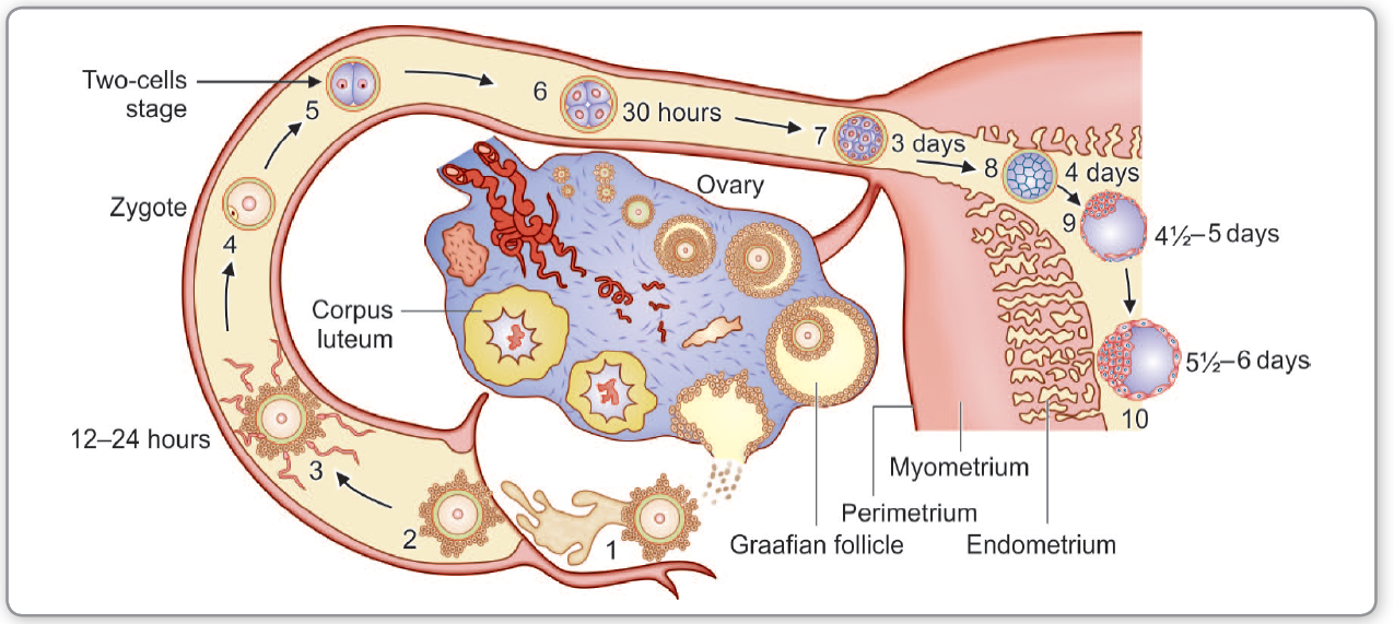
- Definition: implantation outside the uterine cavity. Site: ampulla (most common, ~70%), isthmus, fimbrial, ovarian, cervical, caesarean scar, abdominal.
- Risk factors: PID (Chlamydia/GC) — most important, previous ectopic, tubal surgery, IVF, IUCD in situ, smoking, endometriosis, assisted reproduction.
- Clinical: amenorrhoea + unilateral lower abdominal pain + vaginal bleeding; shoulder-tip pain (diaphragmatic irritation from blood), syncope/collapse = rupture with haemoperitoneum; cervical motion tenderness, adnexal mass/tenderness; may be clinically silent.
- Diagnosis: urine β-hCG (+), TVS (empty uterus, adnexal mass, free fluid), serial serum hCG (slow rise/plateau), progesterone <5 ng/mL (nonviable), laparoscopy (gold standard historically).
- Management:
4. Gestational trophoblastic disease (GTD)
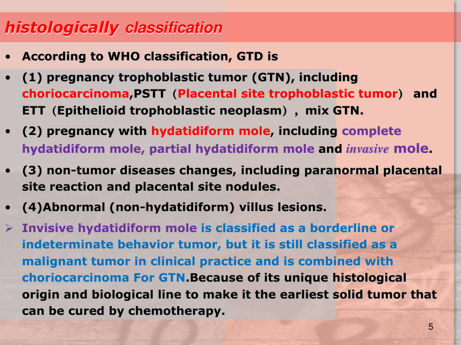
Definition: spectrum of trophoblastic proliferation — hydatidiform mole (complete/partial), invasive mole, choriocarcinoma, PSTT (placental site trophoblastic tumour). Benign mole is premalignant; choriocarcinoma is highly malignant but curable (>90%) with chemotherapy.
| Type | Karyotype | Ultrasound | Malignant potential | hCG |
|---|---|---|---|---|
| Complete mole | 46,XX (all paternal) | "Snowstorm" (vesicular), no fetus, bilateral theca lutein cysts | ~15-20% → invasive mole/GTN | Very high (>100,000) |
| Partial mole | Triploid (69,XXX/XXY) | Fetus present (abnormal), focal molar changes | ~1-5% | Moderately high |
| Invasive mole | As mole | Myometrial invasion | Locally invasive | Elevated, persisting |
| Choriocarcinoma | — | Necrotic/haemorrhagic mass, lung mets | High | Markedly elevated |
Clinical: vaginal bleeding (often 2nd trimester), hyperemesis, preeclampsia <20 weeks (classic clue), hyperthyroidism (hCG cross-reacts with TSH receptor), large-for-dates uterus, grape-like vesicles passed vaginally; theca lutein cysts.
Diagnosis: hCG very high (>100,000), USS snowstorm, histology after evacuation.
Management:
- Surgical evacuation (suction curettage) — the primary treatment for molar pregnancy; under ultrasound guidance; oxytocin after evacuation.
- hCG surveillance is the cornerstone — weekly until negative × 3, then monthly × 6-12 months. Persistent/rising hCG = GTN → treat.
- GTN (invasive mole/choriocarcinoma): chemotherapy — single-agent MTX (low risk) or EMA-CO (high risk); FIGO scoring determines regimen; contraception for 6-12 months (avoid pregnancy until hCG normalised and surveillance complete).
- Follow-up imaging: chest CT (lung mets most common site) if GTN suspected.
High-Yield Points
| Topic | Must-remember |
|---|---|
| First rule | Ectopic until proven otherwise — positive test + pain/bleeding + empty uterus |
| Ectopic site | Ampulla (~70%) most common |
| Ectopic risk | PID (Chlamydia/GC) most important; previous ectopic; IVF |
| Ectopic rupture | Shoulder-tip pain + syncope/collapse = haemoperitoneum emergency |
| MTX criteria | Stable, unruptured, <35 mm, no fetal heartbeat, hCG <5,000 |
| Miscarriage cause | Chromosomal (~50-60%) — most sporadic losses |
| Recurrent loss | Investigate karyotype, cavity, APS antibodies, thyroid, glucose |
| Complete mole | 46,XX all-paternal; snowstorm scan, no fetus; preeclampsia <20 wk |
| Mole hCG | Very high (>100,000); surveillance until negative — cornerstone |
| GTN treatment | Chemotherapy (MTX low-risk; EMA-CO high-risk) |
| Choriocarcinoma | Highly malignant but curable >90% |
| Anti-D | Give to all Rh-negative women with bleeding/evacuation |
Topic Summary
Bleeding in early pregnancy with a positive test requires immediate exclusion of ectopic pregnancy (empty uterus + pain + abnormal hCG → treat as ectopic: MTX or surgery). Miscarriage is managed expectantly, medically, or surgically depending on stage; recurrent loss warrants investigation. GTD spans benign hydatidiform mole to malignant choriocarcinoma: diagnosis by very high hCG + snowstorm scan; treatment by suction evacuation + hCG surveillance; persistent disease = chemotherapy. Anti-D for all Rh-negative women with first-trimester bleeding.
LMCHK OSCE Practice — First-Trimester Bleeding
Station setup: A 29-year-old woman (G1P0) at 8 weeks by LMP presents with painless PV bleeding for 2 days. Urine β-hCG positive. TVS shows a viable intrauterine gestation (CRL 15 mm, fetal heartbeat present) with a small subchorionic haematoma. She is anxious about the bleeding.
Candidate tasks (8 min):
- Take a focused history (bleeding amount, clots, pain, dizziness, past pregnancy loss, Rh status).
- Diagnose threatened miscarriage (viable IUP + bleeding) and explain the likely outcome (many settle; ~50% with bleeding + viable fetus still miscarry; higher if >5 days bleeding).
- Arrange quantitative β-hCG + repeat TVS in 1-2 weeks to confirm ongoing viability.
- Check blood group — if Rh-negative, give Anti-D (300 μg IM).
- Advise: rest, avoid intercourse until bleeding settles, return immediately for heavy bleeding, pain, or fever (infection signs); review for ectopic red flags if pain develops.
Key marking cues:
- Distinguishes threatened (viable IUP) from missed/incomplete/ectopic by scan.
- Anti-D for Rh-negative — mandatory safety point.
- Explains prognosis honestly (bleeding in viable pregnancy = higher loss risk).
- Gives clear red-flag return advice (heavy bleeding, pain, fever, shoulder-tip pain).