Preparatory Mindset
Childhood haematology in this syllabus covers iron-deficiency anaemia (IDA — the most common anaemia of childhood), megaloblastic anaemia (B12/folate), and acute leukaemia (mostly ALL). The mindset:
- Anaemia workup starts with MCV: microcytic (iron deficiency, thalassaemia, chronic disease, sideroblastic) → normocytic (bleeding, haemolysis, aplasia) → macrocytic (B12/folate, hypothyroidism) — with reticulocytes separating production from destruction.
- IDA: microcytic hypochromic, low ferritin, ↑ red cell distribution width, pale child, pica, koilonychia — from inadequate dietary iron (cow's milk <1 yr, exclusive breastfeeding past 6 mo) → oral iron, dietary advice, repeat FBC in 4-6 weeks; refractory → investigate (malabsorption, occult loss, thalassaemia trait).
- Acute leukaemia (ALL most common in children): fever + pallor + bleeding + bone pain + lymphadenopathy/hepatosplenomegaly → pancytopenia + blasts on film → bone marrow aspirate (blast count) is diagnostic → chemotherapy (not surgery); ALL is highly curable (>90% in children).
The exam pearl: "child with pallor + pica" = IDA; "child with bone pain + bruising + fever" = leukaemia.
Core Concepts
1. Anaemia — classification by MCV and reticulocyte
| Type | MCV (fL) | Causes |
|---|---|---|
| Microcytic hypochromic | <80 | Iron deficiency (commonest cause of anaemia overall), thalassaemia, anaemia of chronic disease, sideroblastic |
| Normocytic | 80-100 | Acute bleeding, haemolysis, aplastic/red-cell aplasia, renal disease, chronic infection |
| Macrocytic | >100 | Megaloblastic — B12 deficiency, folate deficiency, drugs (MTX), hypothyroidism, liver disease, erythroleukaemia |
Blood film patterns: IDA — small pale RBCs with increased central pallor, anisopoikilocytosis; megaloblastic — macro-ovalocytes + hypersegmented neutrophils; thalassaemia — target cells, microcytosis with normal RDW.
Initial workup (any anaemia): Hb, Hct, MCV/MCH/MCHC, WBC + differential, platelet count, reticulocyte count, blood film — then targeted tests (ferritin, B12/folate, Hb electrophoresis, DAT, Coombs).
2. Iron-deficiency anaemia (IDA)
Why so common: rapid growth + expanding blood volume; inadequate dietary iron — exclusive breastfeeding past 6 months without weaning iron, cow's milk before 1 year (low iron + poor absorption + occult GI loss), poor weaning solids; premature infants (low stores).
Clinical: pallor, fatigue, irritability, poor appetite, pica (eating dirt/ice), koilonychia (spoon nails), tachycardia, systolic murmur; severe — breathlessness, heart failure; behavioural/cognitive effects (long-term with chronic deficiency).
Investigations:
| Test | IDA finding |
|---|---|
| CBC | Microcytic hypochromic (MCV ↓, MCH ↓), Hb ↓, RDW ↑ |
| Ferritin | Low (<12-15 μg/L) — the best single test (but ↑ in inflammation — check CRP) |
| Serum iron / TIBC | Iron ↓, TIBC ↑, transferrin saturation ↓ |
| Blood film | Hypochromic microcytic, central pallor |
| Stool occult blood | If GI loss suspected (hookworm, malabsorption) |
Management:
- Oral iron (ferrous sulphate 3-6 mg/kg/day elemental) — for 3 months (1 month to normalise Hb + 2 months to replenish stores).
- Dietary advice — iron-rich weaning foods (fortified cereal, meat, egg, legumes), vitamin C enhances absorption (orange juice), avoid tea with meals (tannins ↓ absorption); no cow's milk before 1 year; limit milk after.
- Response check: reticulocyte rise by 3-7 days, Hb rise ≥10-20 g/L in 2-4 weeks — if no response: compliance? wrong diagnosis (thalassaemia trait), ongoing loss (occult GI bleed), malabsorption → investigate.
- Severe/symptomatic or non-absorbing: IV iron; transfusion only if severe/acute compromise.
Differential — thalassaemia trait (common in Chinese/Mediterranean): mild microcytic anaemia, normal/increased ferritin, normal RDW, elevated HbA2 (on electrophoresis); do NOT give iron (no deficiency). This is the classic "IDA that doesn't respond" in Chinese children.
3. Megaloblastic anaemia (B12 / folate deficiency)
- Causes: folate — poor diet, malabsorption, anticonvulsants (phenytoin), haemolytic states; B12 — pernicious anaemia (rare in children), vegan diet, ileal disease (Crohn), congenital B12 defects, fish tapeworm, nitrous oxide.
- Clinical: pallor + glossitis (sore tongue), diarrhoea, neurological (B12 — subacute combined degeneration: paraesthesia, ataxia, vibration loss), mild jaundice (ineffective erythropoiesis).
- Investigations: macrocytic (MCV >100), hypersegmented neutrophils, ↓ B12 or ↓ folate (check both + red cell folate), ↑ LDH, ↑ indirect bilirubin; normal reticulocyte.
- Management: B12 replacement (IM hydroxocobalamin) for B12 deficiency (correct neurological damage); folic acid (oral) for folate deficiency; check B12 BEFORE giving folate alone (folate can mask B12 deficiency and worsen neurology); dietary correction; treat cause.
4. Acute leukaemia (ALL — the paediatric leukaemia)
Epidemiology: ALL is the most common childhood cancer (peak 2-5 years); AML less common (more in adolescents/infants).
Clinical — the "leukaemia syndrome":
- Bone marrow failure: fever (infection), pallor/fatigue (anaemia), bruising/petechiae/bleeding (thrombocytopenia) — the classic triad
- Infiltrative: bone pain/limping (periosteal infiltration — a big clue), lymphadenopathy, hepatosplenomegaly, gum hypertrophy (M4/M5 AML)
- CNS: headache, vomiting, cranial nerve palsy (leptomeningeal)
- Mediastinal mass (T-cell ALL) — superior vena cava syndrome, wheeze
Investigations:
| Test | Finding |
|---|---|
| FBC | Pancytopenia (↓Hb, ↓platelets, ↓WBC or ↑WBC) |
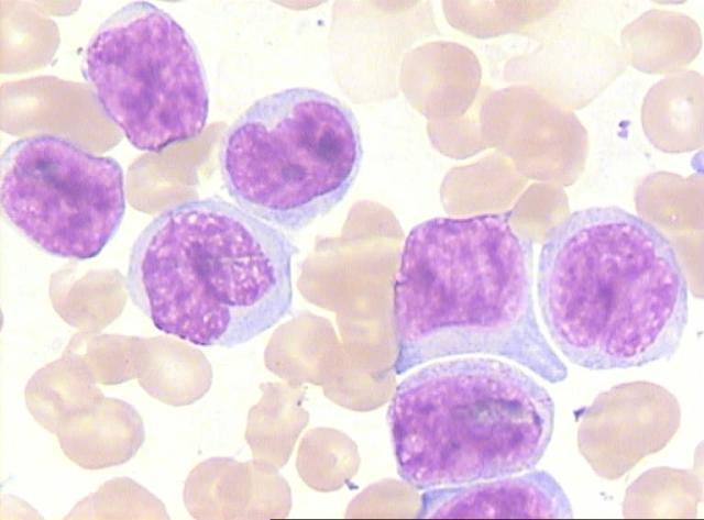
| Blood film | Blasts (immature cells) |
| Bone marrow aspirate + trephine | Diagnostic — blast count >20-25% (ALL: L1/L2/L3 morphology; immunophenotyping — CD markers; cytogenetics — t(12;21), t(9;22) Philadelphia, hyperdiploidy) |
| Lumbar puncture | CNS involvement (blasts in CSF) |
| CXR | Mediastinal mass (T-ALL) |
Management (chemotherapy, NOT surgery):
- Supportive care first: infection prophylaxis (empiric antibiotics for fever + neutropenia), tumour lysis syndrome prevention (hydration, allopurinol/rasburicase, monitor K/PO4/Ca/urate), blood products.
- Multi-phase chemotherapy: induction (remission) → consolidation → maintenance (with intrathecal methotrexate for CNS prophylaxis; cranial irradiation now limited — neurotoxicity).
- High-risk/relapse: allogeneic stem cell transplant (HSCT).
- Prognosis: ALL cure >85-90% in children (favourable: age 1-10, low WBC, hyperdiploidy; unfavourable: infant, high WBC, Philadelphia chromosome, CNS disease).
Complications of treatment: neutropenic sepsis, tumour lysis syndrome, mucositis, cardiomyopathy (anthracyclines), secondary malignancy, growth/endocrine (cranial RT), fertility issues — long-term follow-up.
High-Yield Points
| Topic | Must-remember |
|---|---|
| Anaemia workup | MCV classifies (micro/normo/macrocytic) + reticulocyte |
| IDA | Most common anaemia — microcytic hypochromic, low ferritin, ↑ RDW |
| IDA causes | Cow's milk <1 yr, late weaning, prematurity |
| IDA treatment | Oral iron 3-6 mg/kg/day × 3 months + diet (vitamin C, no tea) |
| IDA response | Hb rise by 2-4 weeks; no response → compliance, thalassaemia trait, occult loss |
| Thalassaemia trait | Microcytic but normal ferritin + ↑ HbA2 — do NOT iron |
| Megaloblastic | MCV >100 + hypersegmented neutrophils |
| B12 vs folate | Check B12 before folate alone (masking) |
| ALL | Most common childhood cancer (2-5 yr) |
| Leukaemia triad | Fever + pallor + bruising/bleeding + bone pain |
| ALL diagnosis | Bone marrow aspirate — blasts >20-25% |
| ALL treatment | Chemotherapy (induction→consolidation→maintenance) + intrathecal MTX |
| ALL prognosis | Cure >85-90% |
| Tumour lysis | Prevent — hydration + rasburicase; monitor K/PO4/Ca/urate |
| AML vs ALL | AML — gum hypertrophy (M4/M5), DIC (M3/PML-RARA) |
Topic Summary
Childhood anaemia is classified by MCV: IDA (microcytic, low ferritin, ↑RDW — treat with oral iron 3 months + diet; thalassaemia trait mimics but has normal ferritin/↑HbA2), megaloblastic (macrocytic, B12/folate — check B12 first), normocytic (bleeding/haemolysis). Acute leukaemia (ALL) presents with fever + pallor + bruising + bone pain + organomegaly; pancytopenia + blasts → bone marrow aspirate (>20-25% blasts) is diagnostic; treatment is chemotherapy (with tumour lysis prevention); childhood ALL is curable in >85-90%.
LMCHK OSCE Practice — Pallor and Bruising in a Toddler
Station setup: A 3-year-old boy presents with 3 weeks of increasing pallor, low-grade fever, and easy bruising. He has been limping (left leg pain at night). Examination: pale, scattered petechiae, cervical lymphadenopathy, liver 3 cm, spleen 4 cm. FBC: Hb 75 g/L, WBC 4.2, platelets 40, blood film shows blasts.
Candidate tasks (8 min):
- Take a focused history (fever, bleeding, bone pain, weight loss, recurrent infections).
- Recognise acute leukaemia (likely ALL) — bone marrow failure (pallor, bruising, fever) + infiltrative (bone pain, lymphadenopathy, hepatosplenomegaly) + blasts on film.
- State the diagnostic test — bone marrow aspirate + trephine (blast count, immunophenotype, cytogenetics) and lumbar puncture (CNS staging).
- Outline immediate management: infection prophylaxis; tumour lysis prevention (hydration, rasburicase, electrolytes); blood products; then chemotherapy (induction → consolidation → maintenance + intrathecal MTX).
- Discuss prognosis honestly (childhood ALL >85-90% cure) and long-term follow-up.
Key marking cues:
- Connects bone pain + bruising + fever = leukaemia until proven otherwise.
- Bone marrow aspirate is the diagnostic test (not just blood film).
- Prevents tumour lysis syndrome.
- Mentions intrathecal chemo (CNS prophylaxis).
- Gives an accurate prognosis and follow-up plan.