Preparatory Mindset
Congenital heart disease (CHD) is the most common congenital malformation (~8/1000) and a guaranteed exam topic. The mindset is the cyanotic vs acyanotic split (determined by the direction of shunt):
- Acyanotic (left-to-right shunt — pulmonary overcirculation): VSD (most common, 30%), ASD, PDA → heart failure, recurrent chest infections, failure to thrive (17CM exam tested); late complications — pulmonary hypertension → Eisenmenger syndrome (shunt reversal → cyanosis) — the reason early repair matters.
- Cyanotic (right-to-left shunt — deoxygenated blood to the systemic circulation): Tetralogy of Fallot (TOF — the "tet spell"), Transposition of the great arteries (TGA), tricuspid atresia, pulmonary atresia → cyanosis, clubbing, polycythaemia; duct-dependent lesions (TGA, pulmonary atresia) need prostaglandin E1 to keep the duct open before surgery.
- The newborn exam: murmur, cyanosis, poor pulses (coarctation), hyperoxia test (does not turn pink in 100% O₂ → cyanotic CHD), pulse oximetry screening at 24 h.
The exam pearl: VSD = the most common CHD; TOF = the most common cyanotic CHD; PDA murmur = continuous "machinery"; TGA = the neonatal emergency (duct-dependent).
Core Concepts
1. Fetal to neonatal circulation (why CHD matters at birth)
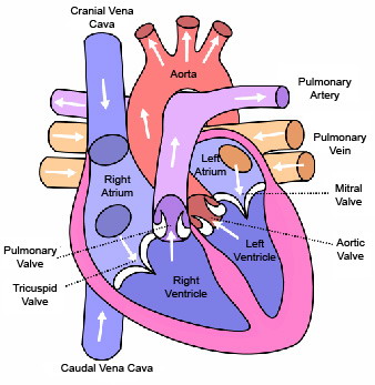
- Fetal: placenta supplies O₂; 3 shunts — ductus venosus, foramen ovale, ductus arteriosus; blood bypasses the lungs.
- At birth: first breaths → pulmonary vascular resistance falls, pulmonary blood flow ↑6-fold; ductus arteriosus closes within hours-days (prostaglandin withdrawal); foramen ovale closes.
- Duct-dependent circulation: some cyanotic lesions (TGA, pulmonary/tricuspid atresia, critical coarctation) require the ductus arteriosus to remain patent to supply pulmonary or systemic blood flow → prostaglandin E1 (alprostadil) infusion keeps the duct open until surgery.
2. Classification of CHD
| Type | Lesions | Direction | Result |
|---|---|---|---|
| Acyanotic (left-to-right shunt) | VSD (30%), ASD (6%), PDA (12%), AVSD | L→R (pulmonary overcirculation) | Heart failure, infections; late Eisenmenger |
| Acyanotic (obstructive) | Aortic stenosis, coarctation of aorta, pulmonary stenosis | No shunt | Pressure overload |
| Cyanotic (right-to-left) | TOF (6%), TGA (5%), tricuspid atresia, pulmonary atresia, total anomalous pulmonary venous drainage | R→L (deoxygenated to body) | Cyanosis, clubbing, polycythaemia |
3. Acyanotic CHD — the left-to-right shunts
| Lesion | Anatomy | Murmur | Clinical |
|---|---|---|---|
| VSD (most common CHD) | Defect in ventricular septum; L→R shunt | Pansystolic at left lower sternal edge (LSE) | Small — asymptomatic; large — heart failure, failure to thrive, recurrent pneumonia; Eisenmenger if unrepaired |
| ASD | Atrial septal defect (secundum most common) | Soft ejection systolic at upper LSE + fixed split S2 | Usually asymptomatic until adult; atrial arrhythmias, paradoxical emboli |
| PDA | Ductus arteriosus remains patent | Continuous "machinery" murmur (left infraclavicular, radiating to back) | Bounding pulses, wide pulse pressure; preterm infants — common (closure with indomethacin/ibuprofen or surgical ligation) |
| AVSD | Endocardial cushion defect (common in Down syndrome) | VSD/ASD murmur mix | Early heart failure |
Heart failure in infants: poor feeding, tachypnoea, sweating with feeds, failure to thrive, hepatomegaly, tachycardia — treat with diuretics (furosemide) + ACE inhibitors + nutritional support; definitive surgical repair.
4. Cyanotic CHD
| Lesion | Anatomy | Features |
|---|---|---|
| TOF (most common cyanotic) | 4 features: VSD + overriding aorta + RV hypertrophy + pulmonary stenosis | Cyanosis (2-6 months onset), squatting, clubbing, "tet spells" (cyanosis crisis — crying/feeding → infundibular spasm → ↓pulmonary flow); pansystolic murmur at LSE; treat spell — knee-chest, O₂, morphine, β-blocker (propranolol), fluid, sodium bicarbonate; surgery — full repair |
| TGA | Aorta arises from RV, pulmonary artery from LV (parallel circulation) | Neonatal emergency — severe cyanosis from birth; duct-dependent — PGE1 infusion + balloon atrial septostomy; arterial switch operation |
| Tricuspid atresia / pulmonary atresia | No tricuspid valve / no pulmonary outflow | Cyanosis at birth; duct-dependent; PGE1; staged surgery (Fontan pathway) |
Cyanosis clues: central cyanosis (lips, tongue) not improved by O₂ (hyperoxia test fails), clubbing (after 3-6 months), polycythaemia (↑ Hb from chronic hypoxia), squatting (TOF — increases systemic resistance → ↑ pulmonary flow), syncope/cyanotic spells.
Tet spell management (the exam drill): knee-chest position (↑ SVR) + 100% O₂ + morphine (↓ infundibular spasm) + IV fluid bolus + IV propranolol (β-blocker); sodium bicarbonate for acidosis — then urgent surgery (BT shunt or full repair).
5. Coarctation of aorta (acyanotic, obstructive)
- Narrowing of the aorta (usually near ductus) — upper limb hypertension, weak/delayed femoral pulses, radiofemoral delay, murmur (posterior interscapular).
- Infantile (critical) form: presents with heart failure/shock when duct closes — PGE1 + surgery.
- Associated with Turner syndrome, bicuspid aortic valve.
- Treatment — surgical resection ± balloon angioplasty; long-term hypertension follow-up.
6. Murmurs — innocent vs pathological
Innocent murmur — the "five Ss": Systolic, Soft, Short, Small area, Symptoms absent (plus: changes with position, no radiation, normal heart sounds, child thriving).
Features of pathological murmur: diastolic, pansystolic, loud (>3/6), radiating, fixed split S2, associated cyanosis/weak pulses/heart failure/poor growth.
Evaluation: examination (femoral pulses, BP 4 limbs, SpO₂) → ECHO (the diagnostic test); CXR (cardiomegaly, pulmonary plethora/oligemia), ECG; pulse oximetry screening in newborns.

High-Yield Points
| Topic | Must-remember |
|---|---|
| Most common CHD | VSD (30%) |
| Most common cyanotic | TOF (6%) |
| PDA murmur | Continuous "machinery" |
| VSD murmur | Pansystolic at LSE |
| ASD murmur | Ejection systolic + fixed split S2 |
| TOF tetrad | VSD + overriding aorta + RVH + PS |
| Tet spell treatment | Knee-chest + O₂ + morphine + β-blocker + fluids |
| TGA | Neonatal emergency — cyanosis at birth; duct-dependent (PGE1) |
| Duct-dependent lesions | TGA, pulmonary/tricuspid atresia — PGE1 infusion |
| Coarctation | Weak femoral pulses + radiofemoral delay + upper limb HTN |
| Infant heart failure | Poor feeding, tachypnoea, sweating, FTT, hepatomegaly |
| AVSD | Down syndrome association |
| PDA in preterm | Close with indomethacin/ibuprofen |
| Hyperoxia test | Cyanosis not improved by 100% O₂ → cyanotic CHD |
| Innocent murmur | 5 Ss — systolic, soft, short, small area, symptoms absent |
| Diagnosis | ECHO is the diagnostic test |
Topic Summary
CHD is classified by shunt direction: acyanotic (VSD most common, ASD, PDA — left-to-right, heart failure/infections; late Eisenmenger if unrepaired) vs cyanotic (TOF most common, TGA — right-to-left, cyanosis/clubbing). TOF tet spells are managed with knee-chest + O₂ + morphine + β-blocker; TGA and other duct-dependent lesions need PGE1 before surgery; coarctation shows weak femoral pulses + upper limb hypertension. Heart failure in infants (poor feeding, sweating, FTT) is treated medically then surgically; ECHO is the diagnostic test; innocent murmurs satisfy the five Ss.
LMCHK OSCE Practice — The Cyanotic Newborn
Station setup: A term newborn is noted to be blue at 6 hours of age, with poor feeding. Pulse oximetry: preductal SpO₂ 88%, postductal 75%. Heart: loud single S2, no murmur. Chest clear. On 100% O₂, SpO₂ improves only to 90%.
Candidate tasks (8 min):
- Recognise central cyanosis with a hyperoxia test that does NOT normalise → cyanotic CHD (postductal < preductal → suggests duct-dependent lesion).
- List differentials: TGA (most likely in a neonate with loud single S2), TOF, pulmonary atresia, tricuspid atresia (plus sepsis/PPHN — always consider).
- Order urgent investigations: ECHO (the diagnostic test), blood gas, CXR, glucose/calcium/sepsis screen.
- Start PGE1 (alprostadil) infusion immediately (duct-dependent circulation — before ECHO if suspected) + prostaglandin side effects: apnoea (be ready to ventilate), fever, hypotension.
- Discuss definitive management: e.g., TGA → arterial switch operation; transfer to cardiac centre; support parents.
Key marking cues:
- Hyperoxia test interpretation — cyanosis not corrected = structural CHD.
- PGE1 started early (duct-dependent) — the life-saving step.
- Orders ECHO as the diagnostic test.
- Knows PGE1 side effects (apnoea) and prepares ventilation.
- Plans urgent transfer to paediatric cardiology.