# Ch01: Introduction — Concepts, Layers & Basic Structures
Preparatory Mindset
Regional (topographical) anatomy studies the body by regions (head, neck, trunk, limbs), focusing on:
- Relationships of visceral organs, muscles, nerves and vessels within each region
- Layers of each body division (skin → fascia → muscle → bone)
- Clinical application — where to puncture, incise, palpate, and which structures are at risk
Exam mindset: You are not just memorizing lists — you are building a 3-D mental model. Every structure has a location, a layer, a blood supply, an innervation, and a clinical consequence. The 局解 exam rewards students who can answer "what is at risk if I cut here?" and "which nerve/vessel is compressed by this hernia/fracture?"
Core Concepts
1. Divisions of the body (based on systematic anatomy)
- Head → scalp/face; Neck → anterior/posterior triangles + root; Trunk → thorax, abdomen, pelvis; Limbs → upper (shoulder/arm/forearm/hand) & lower (gluteal/thigh/leg/foot)
2. Layers of the body (from superficial to deep) — MUST KNOW
| Layer | Contents / Notes |
|---|---|
| Skin | Epidermis + dermis; thickness 1–4 mm; follows Langer's lines (incisions parallel to these heal with less scarring) |
| Superficial fascia (subcutaneous) | Loose CT + fat; contains superficial arteries/veins, cutaneous nerves, lymphatics & nodes |
| Deep fascia (proper fascia) | Dense CT; contains deep arteries, veins, nerves; forms specialized structures |
| Muscles | Skeletal muscle in compartments |
| Bones & joints | Deepest layer; provide support |
3. Structures formed by deep fascia (must-know list)
- Intermuscular septum: deep fascia extends between muscles, attaches to bone → separates muscle groups
- Osseofascial sheath: intermuscular septa + deep fascia + bone enclose a muscle group (e.g., thigh compartments)
- Neurovascular sheath: fascia around a neurovascular bundle (e.g., carotid sheath, axillary sheath)
- Retinaculum: thickened deep fascia that holds tendons in place across joints (e.g., flexor/extensor retinacula)
4. How to learn clinically
- For every region ask: boundaries → layers → contents → relationships → clinical relevance
- Key clinical examples raised in lecture: femoral vein puncture (femoral triangle), cubital fossa (arterial pulse / venipuncture), surgical approaches respecting vessels & nerves
High-Yield Points
- Mnemonic for layers of a limb cross-section (superficial → deep): "Skin → Super-Fat → Deep-Fascia → Muscle → Bone"
- Langer's lines: incisions along cleavage lines → minimal scarring; face incisions follow natural lines
- Deep fascia specializations: septum (separates), sheath (encloses neurovascular), retinaculum (restrains tendons) — know one example of each
- Superficial vs deep fascia: superficial = loose + fat + superficial vessels/nerves/lymph; deep = dense + deep vessels/nerves; the plane of dissection differs
- Scalp = special case: 5 layers (see Head & Neck chapter) — the first 3 are tightly bound as "scalp proper"
Topic Summary
Regional anatomy = structure + relationships + layers + clinical application. The fundamental skill is identifying layers from skin to bone and knowing what runs in each layer (superficial fascia: superficial vessels/nerves/lymphatics; deep fascia: deep neurovascular structures + its specialized formations: intermuscular septum, osseofascial sheath, neurovascular sheath, retinaculum). Every subsequent chapter (limbs, head & neck, thorax, abdomen, pelvis) is built on this layer concept.
LMCHK OSCE Practice
- Surface anatomy check: can you palpate the key bony landmarks (ASIS, pubic tubercle, patella, malleoli, epicondyles, styloid processes) and predict what lies beneath?
- Femoral vein puncture (central line / blood sampling): needle is placed medial to the femoral artery pulse, in the femoral triangle — be able to describe the layers pierced (skin → superficial fascia → deep fascia → femoral sheath).
- Cubital fossa venipuncture: median cubital vein lies superficial to the bicipital aponeurosis — the aponeurosis protects the brachial artery & median nerve beneath.
Illustrations(图解速览)
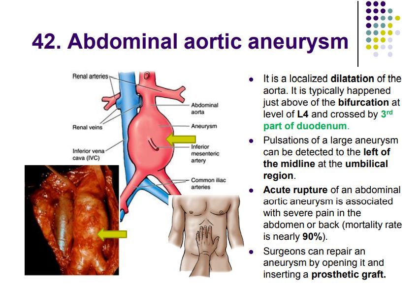
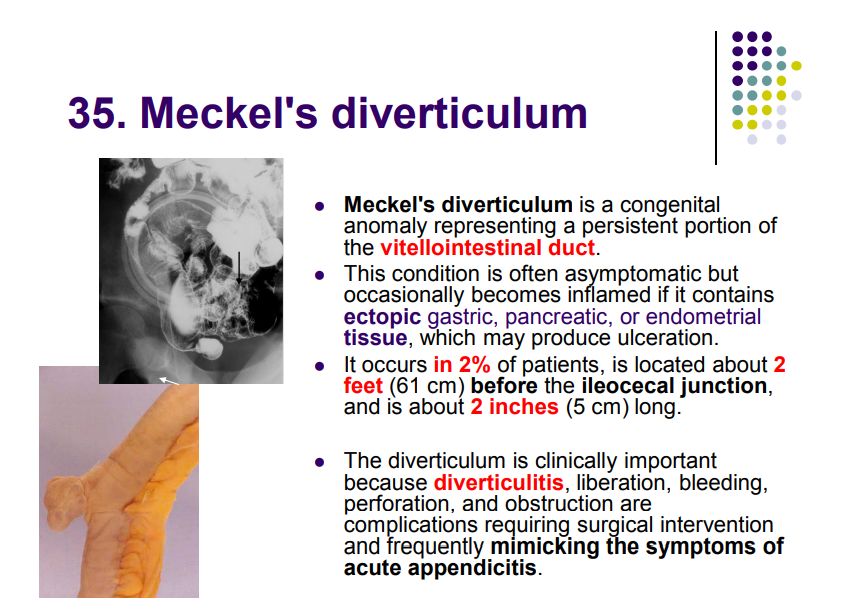
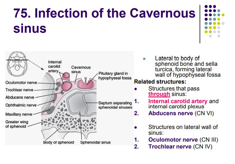
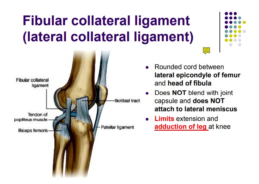
Regional anatomy diagrams — identify the structures shown with the chapter content: