# Ch11: Scalp & Face (SCALP, Cavernous Sinus, Parotid)
Preparatory Mindset
The scalp's five layers and the face's facial nerve / parotid gland relationships are the core. The cavernous sinus (with its structures passing through vs on the lateral wall) is a guaranteed exam question, and the dangerous area of the scalp (loose areolar layer) explains infection spread.
Exam mindset: "Analyze 'SCALP' and its clinical application" and "Analyze the relation between the facial nerve and parotid gland" are the lecture's stated skill objectives. Know the five layers, the three neurovascular groups, the cavernous sinus content list, and the parotid's longitudinal vs transverse structures.
Core Concepts
1. Fronto-parieto-occipital region (scalp proper)
1. Skin — thick, greatest concentration of hair & sebaceous glands 2. Connective tissue (superficial fascia) — dense, binds skin to aponeurosis; vessels & nerves run here; rich anastomosis → wounds bleed profusely but heal well 3. Aponeurosis (galea aponeurotica) — tough, connects frontal & occipital bellies of occipitofrontalis; deep transverse lacerations gape widely (must suture the aponeurosis) 4. Loose areolar tissue (subaponeurotic space) — the "dangerous area": contains emissary veins (communicate with intracranial venous sinuses); allows the scalp to move; hematoma spreads widely (to eyelids → "black eye") 5. Pericranium (periosteum) — fuses firmly at sutures; limits subperiosteal hematoma to one bone
- Layers 1-3 are tightly bound = "scalp proper" - Lacerations: bleeding profusely (anastomoses) but heal well; suture the aponeurosis even if skin appears intact - Subaponeurotic hematoma → spreads over the whole calvaria + black eye (blood tracks to eyelids) - Subperiosteal hematoma → limited to one bone (suture boundaries) - Dangerous area infection → can spread via emissary veins to intracranial sinuses (meningitis, cavernous sinus thrombosis) - Incisions should be radiating with the flap pedicle at the base (vessels run from periphery to vertex)
- Boundaries: anterior = supraorbital margin; posterior = external occipital protuberance & superior nuchal line; lateral = superior temporal line
- Five layers (superficial → deep) — mnemonic "SCALP":
- Clinical:
2. Vessels & nerves of the scalp (3 groups)
| Group | Nerves | Vessels |
|---|---|---|
| Anterior | Supratrochlear & supraorbital nerves (V1) | Supratrochlear & supraorbital arteries (from ophthalmic a.) |
| Middle | Auriculotemporal n. (V3) in front of auricle; lesser occipital n. (C2) behind | Superficial temporal a.; posterior auricular a. |
| Posterior | Greater occipital n. (C2) | Occipital a. |
- Clinical: the vessels/nerves enter from the periphery → incisions with pedicle at the base preserve the flap; the superficial temporal artery (in front of the auricle) is used for temporal artery biopsy (giant cell arteritis)
3. Temporal region
- Boundaries: superior temporal line / zygomatic arch / junction of frontal & zygomatic bones / posterior superior temporal line
- Layers: skin → superficial fascia (superficial temporal a. & v., auriculotemporal n.; posterior auricular a. & v., lesser occipital n.) → temporal fascia → temporalis → periosteum
- Clinical: pterion (junction of frontal, parietal, temporal, sphenoid) overlies the middle meningeal artery → fracture here → epidural hematoma (concept 74)
4. Face — superficial structures
- Skin: thin, soft, follows cleavage lines (incisions along these lines heal well)
- Superficial fascia: loose CT, little fat, many vessels/lymphatics → edema accumulates here (renal/cardiac disease → eyelid edema)
- Facial muscles (muscles of expression): orbicularis oculi, orbicularis oris, buccinator, levator anguli oris/labii superioris, risorius, depressor labii/oris, nasalis, platysma — all supplied by the facial nerve (CN VII)
- No deep fascia in the face (skin connected to bones by loose CT)
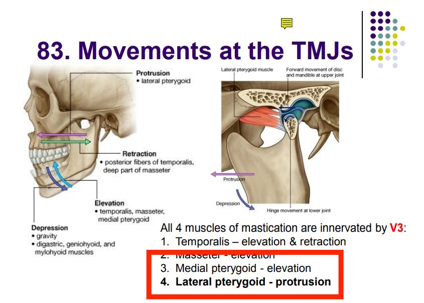
- Masticatory muscles: masseter (elevates), temporalis (elevates & retracts), medial & lateral pterygoids (elevation/protrusion/side-to-side) — all supplied by CN V3 (mandibular division)
5. Facial nerve (CN VII) — concept 78
- Course: leaves the skull through the internal acoustic meatus → facial canal → stylomastoid foramen; enters the parotid gland; divides into five terminal branches
- Five terminal branches (mnemonic "Two Zebras Bit My Cat"): Temporal, Zygomatic, Buccal, Marginal mandibular, Cervical
- Functions: motor to facial muscles; taste to anterior 2/3 of tongue (chorda tympani); parasympathetic (secretomotor) to submandibular/sublingual/lacrimal glands
- Clinical: Bell's palsy (LMN lesion) → whole half of face paralyzed; UMN lesion (stroke) → contralateral lower face only (upper face spared — frontalis & orbicularis oculi have bilateral UMN supply); parotid tumor → facial paralysis
6. Parotid gland (MUST KNOW)
- Longitudinal (vertical): external carotid artery (→ superficial temporal & maxillary), retromandibular vein, auriculotemporal nerve - Transversal (horizontal): transverse facial artery, branches of the facial nerve (parotid plexus) - Mnemonic for vertical: "E-R-A-T" (External carotid, Retromandibular vein, Auriculotemporal n.)... plus superficial temporal
- Position: below the external auditory meatus, behind the ramus of the mandible, in front of SCM; divided by the facial nerve into superficial & deep parts (isthmus between)
- Parotid duct (Stensen's duct): emerges from the anterior border, runs over masseter (1.5 cm below the zygomatic arch), pierces buccinator, opens opposite the second upper molar
- Structures passing through the gland:
- Relations ("parotid bed"): internal carotid a., internal jugular v., CN IX-XII, styloid process — posteromedial
- Clinical: mumps (parotitis) → painful swelling; parotid tumor may compress the facial nerve → facial paralysis; parotidectomy must preserve the facial nerve (parotid plexus makes surgery difficult)
7. Cavernous sinus (concept 75 — MUST KNOW)
1. Internal carotid artery (+ internal carotid sympathetic plexus) 2. Abducens nerve (CN VI) — the only cranial nerve INSIDE the sinus
1. Oculomotor nerve (CN III) 2. Trochlear nerve (CN IV) 3. Ophthalmic nerve (V1) 4. Maxillary nerve (V2)
- Location: lateral to the body of the sphenoid bone and the sella turcica, forming the lateral wall of the hypophyseal fossa
- Structures passing THROUGH the sinus (in the blood):
- Structures in the LATERAL WALL of the sinus (from above):
- Mnemonic: "O T O M" (lateral wall from top): Oculomotor, Trochlear, Ophthalmic, Maxillary — "OTOM" or "O T O M" (the 'M' is V2)
- Communications: connects with the facial vein (via ophthalmic veins & pterygoid plexus) → danger triangle of the face (infections around nose/upper lip can spread to the cavernous sinus → cavernous sinus thrombosis: proptosis, chemosis, CN III/IV/V1/V2/VI palsies, meningitis)
- Clinical: internal carotid aneurysm in the sinus → CN VI palsy (diplopia); pituitary tumors (sella) compress lateral wall structures
High-Yield Points
- SCALP layers: Skin, Connective tissue, Aponeurosis, Loose areolar (dangerous area), Pericranium
- Scalp vessels/nerves: anterior (supratrochlear/supraorbital), middle (auriculotemporal/superficial temporal), posterior (greater occipital/occipital)
- Cavernous sinus: THROUGH = ICA + CN VI; LATERAL WALL = CN III, IV, V1, V2 ("OTOM")
- Parotid: longitudinal = external carotid, retromandibular vein, auriculotemporal n.; transversal = facial n. branches, transverse facial a.; duct → opposite 2nd upper molar
- Facial nerve branches: "Two Zebras Bit My Cat" (Temporal, Zygomatic, Buccal, Marginal mandibular, Cervical)
- Bell's palsy (LMN): whole face; stroke (UMN): contralateral lower face
- Pterion → middle meningeal artery → epidural hematoma (lens-shaped)
- Danger triangle of the face → cavernous sinus thrombosis (via facial→ophthalmic veins)
Topic Summary
The scalp has five layers (SCALP; loose areolar layer = dangerous area with emissary veins → intracranial spread). Its vessels/nerves come in three groups from the periphery. The face has no deep fascia; the facial muscles (CN VII) lie in loose superficial fascia. The parotid gland is crossed longitudinally by the external carotid, retromandibular vein, auriculotemporal nerve, and transversely by facial nerve branches & the transverse facial artery; its duct opens at the 2nd upper molar. The cavernous sinus carries the ICA & CN VI through it and CN III, IV, V1, V2 in its lateral wall — infections from the face's danger triangle can thrombose it.
LMCHK OSCE Practice
- Cranial nerve exam (face): test CN V (sensation, masseter) & CN VII (facial movements: raise eyebrows, close eyes tight, show teeth, puff cheeks); distinguish UMN vs LMN facial weakness.
- Parotid palpation & duct: inspect the parotid region; locate the parotid duct opening opposite the 2nd upper molar (Stensen's duct).
- Temporal artery palpation: for giant cell arteritis — describe the superficial temporal artery course.
- Epidural hematoma scenario: pterion fracture → middle meningeal artery tear → lucid interval → lentiform bleed on CT; urgent evacuation.
- Cavernous sinus thrombosis scenario: facial furuncle + proptosis, chemosis, ophthalmoplegia — explain the emissary/venous pathway.
- Bell's palsy vs stroke: LMN (whole side, forehead involved) vs UMN (lower face only, forehead spared).
Illustrations(图解速览)
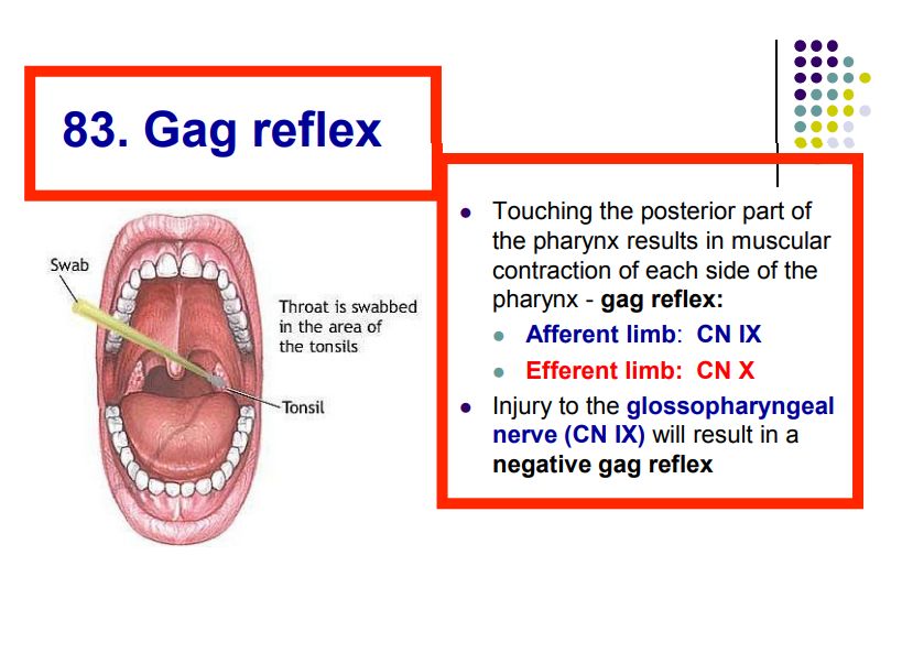
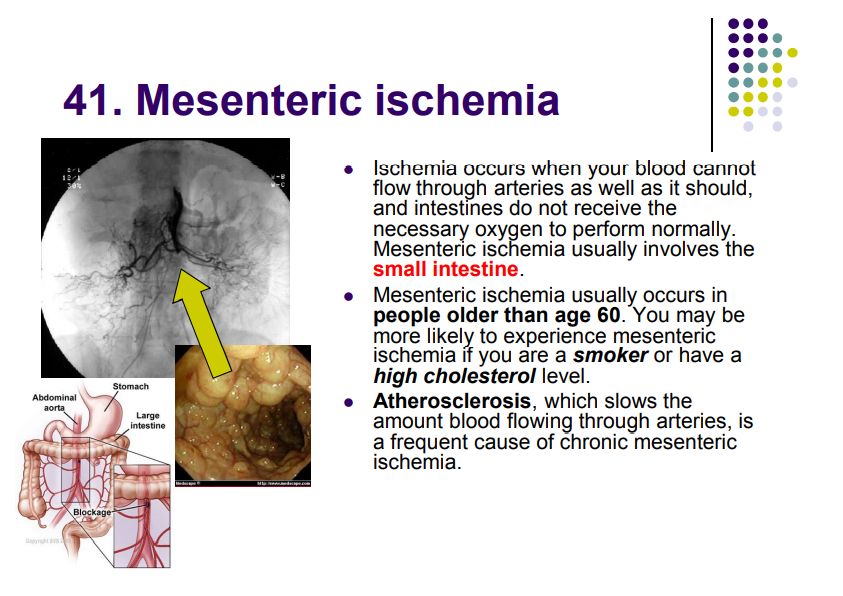
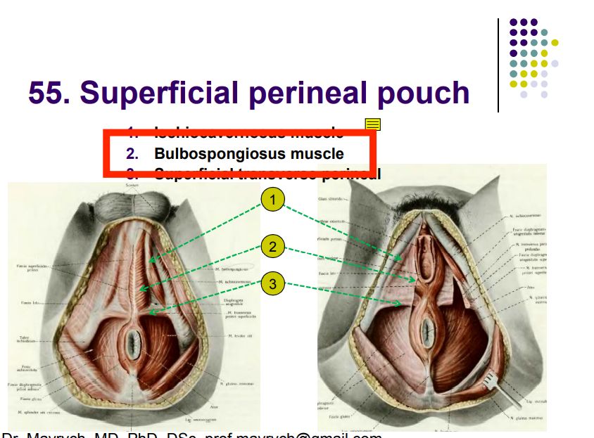
Regional anatomy diagrams — identify the structures shown with the chapter content: