# Ch16: Abdomen I — Anterolateral Abdominal Wall & Inguinal Region
Preparatory Mindset
The abdominal wall is the layer model applied to the trunk: skin → superficial fascia (Camper's & Scarpa's) → muscles (external oblique, internal oblique, transversus) → transversalis fascia → peritoneum. The inguinal canal is the single most important structure — the site of inguinal hernias (direct vs indirect), the lecture's key/difficult point. The Hesselbach (inguinal) triangle distinguishes them.
Exam mindset: "Layers of the anterolateral abdominal wall", "walls & contents of the inguinal canal", "boundaries of the inguinal triangle", and "direct vs indirect hernia" are guaranteed. The 局解mc大全 is full of these (MCQ: 45-year-old man post-appendectomy develops direct inguinal hernia; 15-year-old boy with left indirect hernia causing obstruction; finger invagination through the superficial ring during exam).
Core Concepts
1. Layers of the anterolateral abdominal wall (MUST KNOW)
From superficial to deep:
- Camper's fascia (superficial fatty layer) - Scarpa's fascia (deep membranous layer) — continues into the thigh as fascia lata (attached below the inguinal ligament); in the scrotum it becomes the dartos fascia; in the penis, the superficial perineal (Colles') fascia - Clinical: rupture of the urethra → urine extravasation follows Scarpa's/Colles' fascial planes (scrotum, penis, anterior abdominal wall — NOT the thigh, because Scarpa's attaches to fascia lata below the inguinal ligament)
- External oblique — aponeurosis forms the inguinal ligament (ASIS → pubic tubercle), the superficial inguinal ring (triangular hiatus), and the lacunar & pectineal ligaments; contributes to the anterior wall of the rectus sheath - Internal oblique — its aponeurosis splits around the rectus (anterior & posterior layers); its lower fibers form the conjoint (falx) tendon with transversus; gives rise to the cremaster muscle (around the spermatic cord) - Transversus abdominis — aponeurosis contributes to the posterior rectus sheath above the arcuate line
- Skin
- Superficial fascia:
- Deep fascia (thin on the abdomen)
- Muscles:
- Transversalis fascia — lines the deep surface; forms the deep inguinal ring and the internal spermatic fascia
- Extraperitoneal fat
- Parietal peritoneum
2. Rectus sheath & linea alba
- Rectus abdominis enclosed in the rectus sheath, formed by the aponeuroses of the three flat muscles
- Above the arcuate line (approximately halfway between umbilicus & pubic symphysis): anterior wall = external oblique + anterior layer of internal oblique; posterior wall = posterior layer of internal oblique + transversus
- Below the arcuate line: ALL aponeuroses pass in front → posterior wall is deficient (only transversalis fascia) — the arcuate line is the transition
- Linea alba: midline fusion of aponeuroses — avascular → midline (median) incision is preferred (minimal bleeding); also site of linea alba hernia
- Pyramidalis (small, inconstant) in front of the lower rectus
3. Blood supply & innervation of the wall
- Arteries: superior epigastric (from internal thoracic), inferior epigastric (from external iliac — crosses lateral to the deep inguinal ring), superficial epigastric, deep & superficial circumflex iliac, posterior intercostal/lumbar
- Nerves: lower 6 thoracic nerves (T7-T12), iliohypogastric & ilioinguinal nerves (L1), genitofemoral (L1-2)
- Clinical: the inferior epigastric artery forms the lateral border of the Hesselbach triangle (landmark for direct vs indirect hernia); pain from the diaphragm (C3-5) may be referred to the shoulder, not the abdominal wall
4. Inguinal canal (MUST KNOW — the lecture's key point)
- Anterior: external oblique aponeurosis (lateral 1/3 also internal oblique) - Posterior: transversalis fascia (medially reinforced by the conjoint tendon) - Floor: inguinal ligament (medially the lacunar ligament) - Roof: internal oblique & transversus (arch over the canal)
- Male: spermatic cord (ductus deferens, testicular artery, pampiniform plexus, genital branch of genitofemoral nerve, lymphatics) + ilioinguinal nerve (superficial) - Female: round ligament of the uterus + ilioinguinal nerve
- Position: oblique passage through the lower abdominal wall, above & parallel to the medial half of the inguinal ligament, from the deep ring (lateral) to the superficial ring (medial), ~4 cm long
- Deep (internal) inguinal ring: opening in the transversalis fascia, lateral to the inferior epigastric vessels, above the midpoint of the inguinal ligament
- Superficial (external) inguinal ring: triangular hiatus in the external oblique aponeurosis, medial to the inferior epigastric vessels, above & lateral to the pubic tubercle
- Walls:
- Contents:
- Fascial coverings of the cord (inside out): internal spermatic fascia (transversalis), cremasteric fascia (internal oblique), external spermatic fascia (external oblique aponeurosis)
5. Hesselbach (inguinal) triangle — MUST KNOW
- Boundaries: medial = lateral border of rectus abdominis; lateral = inferior epigastric vessels; inferior = inguinal ligament (medial half)
- Significance: the site where direct inguinal hernias emerge (medial to the inferior epigastric vessels, through the posterior wall)
- Indirect inguinal hernias enter through the deep ring, lateral to the inferior epigastric vessels, pass through the canal, may descend into the scrotum
- MCQ answers (局解mc大全): indirect = lateral to inferior epigastric vessels, congenital (patent processus vaginalis), may reach the scrotum; direct = medial to inferior epigastric vessels (Hesselbach triangle), acquired (weak posterior wall), rarely enters the scrotum, often in older men after strain
6. Inguinal hernia — clinical pearls
| Feature | Indirect | Direct |
|---|---|---|
| Entry | Deep ring (lateral to inf. epigastric a.) | Hesselbach triangle (medial to inf. epigastric a.) |
| Age | Congenital/young (patent processus vaginalis) | Acquired/older (weak wall) |
| Course | Through the canal, may reach scrotum | Pushes directly forward through the posterior wall |
| Reduction | Reducible unless strangulated; finger pressure on the deep ring controls it | Bulges forward; pressure on the deep ring does NOT control it |
| Strangulation risk | Higher (narrow neck at deep ring) | Lower (wide neck) |
- Clinical tests: invaginate the scrotal skin to palpate the superficial ring; reduce the hernia & press the deep ring → if it stays reduced = indirect
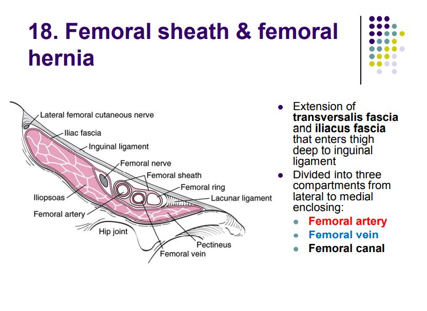
- Femoral hernia (recap): below & lateral to the pubic tubercle, more common in women, high strangulation (lacunar ligament)
- MCQ: 45-year-old man after appendectomy develops a direct inguinal hernia (weak posterior wall, medial to epigastric vessels); 15-year-old boy with obstruction → indirect hernia (patent processus vaginalis)
High-Yield Points
- Wall layers mnemonic: "Skin, Camper, Scarpa, External, Internal, Transversus, Transversalis, Peritoneum" (SCIETTTP) — know Camper (fatty) vs Scarpa (membranous)
- Scarpa's fascia → fascia lata (thigh), dartos (scrotum), Colles' (perineum); urine extravasation follows these planes
- Rectus sheath: above arcuate line, posterior wall = internal oblique + transversus; below = all aponeuroses in front (posterior wall = transversalis only)
- Inguinal canal: deep ring lateral to inferior epigastric a.; superficial ring medial to it
- Indirect = lateral to inferior epigastric (deep ring), congenital, may reach scrotum; Direct = medial (Hesselbach triangle), acquired
- Hesselbach triangle: medial = rectus, lateral = inferior epigastric a., inferior = inguinal ligament
- Cord coverings: "I-C-E" (Internal spermatic, Cremasteric, External spermatic) from deep ring out
- Linea alba = avascular → midline incisions
- Cremaster muscle = from internal oblique (genital branch of genitofemoral nerve → cremasteric reflex)
Topic Summary
The anterolateral abdominal wall has 7 layers (skin, Camper's, Scarpa's, external oblique, internal oblique, transversus, transversalis + peritoneum); the rectus sheath changes at the arcuate line; the linea alba is the avascular midline. The inguinal canal runs from the deep ring (lateral to inferior epigastric vessels) to the superficial ring (medial), with walls anterior/posterior/floor/roof and the spermatic cord (male) or round ligament (female) as contents. The Hesselbach triangle (rectus medial, inferior epigastric lateral, inguinal ligament inferior) is where direct hernias emerge — medial to the inferior epigastric vessels — while indirect hernias enter the deep ring lateral to those vessels. This distinction is the classic 局解 exam question.
LMCHK OSCE Practice
- Hernia examination (standing then supine): inspect both groins & scrotum; reduce the hernia; locate the deep ring; invaginate the superficial ring; ask the patient to cough (increased abdominal pressure).
- Direct vs indirect: reduce the hernia, press firmly over the deep ring (midpoint of inguinal ligament) — if it stays reduced with coughing = indirect; if it bulges = direct (posterior wall defect).
- Cremasteric reflex: stroke the medial thigh → ipsilateral scrotum elevates (ilioinguinal/genitofemoral nerve); test in spinal cord injury assessment.
- Urinary retention / suprapubic catheter: the bladder is extraperitoneal behind the pubic symphysis; describe the midline suprapubic approach through the linea alba.
- Abdominal incisions: midline (linea alba — avascular), transverse (McBurney for appendectomy), paramedian — explain the layer anatomy for each.
- Appendicitis pain (concept 37): visceral (umbilical) → somatic (right iliac fossa) migration — describe the McBurney point (1/3 of the line from ASIS to umbilicus).
Illustrations(图解速览)
Regional anatomy diagrams — identify the structures shown with the chapter content: