# Ch07: Upper Limb I — Pectoral Region & Axilla
Preparatory Mindset
The axilla is the "gateway" of the upper limb — a pyramidal space containing the axillary vessels, the brachial plexus, and the axillary lymph nodes. The pectoral region includes the breast and the pectoral muscles.
Exam mindset: The lecture flags these MUST-KNOW items: (1) contents of the axilla, (2) branches of the axillary artery, (3) main branches (six) of the brachial plexus. The breast (lymphatic drainage, concept 21) and axillary lymph node groups are classic MCQ topics (局解mc大全: "In the lymphatic drainage of the breast, most (~75%) eventually drains into which group of nodes?").
Core Concepts
1. Pectoral region — layers & muscles
- Layers: skin → superficial fascia (breast in female) → deep fascia (clavipectoral fascia) → muscles → ribs/intercostals
- Clavipectoral fascia (deep to pectoralis major, spans between pectoralis minor, subclavius & coracoid process): pierced by the cephalic vein, thoracoacromial vessels, and lateral pectoral nerve
- Muscles: pectoralis major, pectoralis minor, subclavius, serratus anterior
2. Breast (concept 21)
- ~75% → axillary lymph nodes (anterior/pectoral group first, then central → apical → supraclavicular) - Medial part → internal thoracic (parasternal) nodes - Cross-drainage to the opposite breast
- Position: over the pectoral fascia & pectoralis major, between 2nd–6th ribs, sternum to midaxillary line; tail of Spence extends to the axilla
- Structure: 15–20 lactiferous lobes → lactiferous ducts → ampulla → open at the nipple; supported by suspensory (Cooper's) ligaments
- Blood supply: internal thoracic (internal mammary) a., lateral thoracic a., intercostal arteries
- Lymphatic drainage (MUST KNOW):
- Clinical: breast cancer spread via axillary nodes; sentinel lymph node biopsy; peau d'orange (Cooper's ligament infiltration); retraction of nipple
3. Axilla
- Apex: cervico-axillary canal (between clavicle, 1st rib, scapula) — where axillary vessels & brachial plexus enter from the neck - Base: skin & fascia of the axilla (axillary fascia) - Anterior wall: pectoralis major & minor, clavipectoral fascia - Posterior wall: latissimus dorsi, teres major, subscapularis - Medial wall: serratus anterior + thoracic wall (ribs 1–4) - Lateral wall: intertubercular (bicipital) groove of the humerus
- Shape: pyramidal space at the junction of arm & thorax
- Boundaries:
- Contents: axillary artery & vein, brachial plexus (cords & branches), axillary lymph nodes, fat, long thoracic nerve (on serratus anterior), thoracodorsal nerve (to latissimus dorsi)
4. Axillary artery (branches — MUST KNOW)
- Part 1 (medial to pec minor): Superior thoracic artery (1 branch) - Part 2 (behind pec minor): Thoracoacromial artery + Lateral thoracic artery (2 branches) - Part 3 (lateral to pec minor): Subscapular artery, Anterior humeral circumflex, Posterior humeral circumflex (3 branches)
- Continuation of the subclavian artery, from the lateral border of the 1st rib to the inferior border of teres major (becomes brachial artery)
- Divided into 3 parts by pectoralis minor:
- Mnemonic: "Screw The Lawyer, Save A Patient" = Superior thoracic, Thoracoacromial, Lateral thoracic, Subscapular, Anterior & Posterior humeral circumflex
- Clinical: axillary artery can be compressed against the humeral head; axillary artery aneurysm/thoracic outlet syndrome
5. Axillary vein
- Formed by the union of basilic vein & brachial veins at the inferior border of teres major
- Runs medial to the artery; receives cephalic vein at the upper part; becomes subclavian vein at the lateral border of the 1st rib
- Clinical: axillary vein thrombosis (Paget–Schroetter, effort thrombosis); central line access
6. Brachial plexus (six main branches — MUST KNOW)
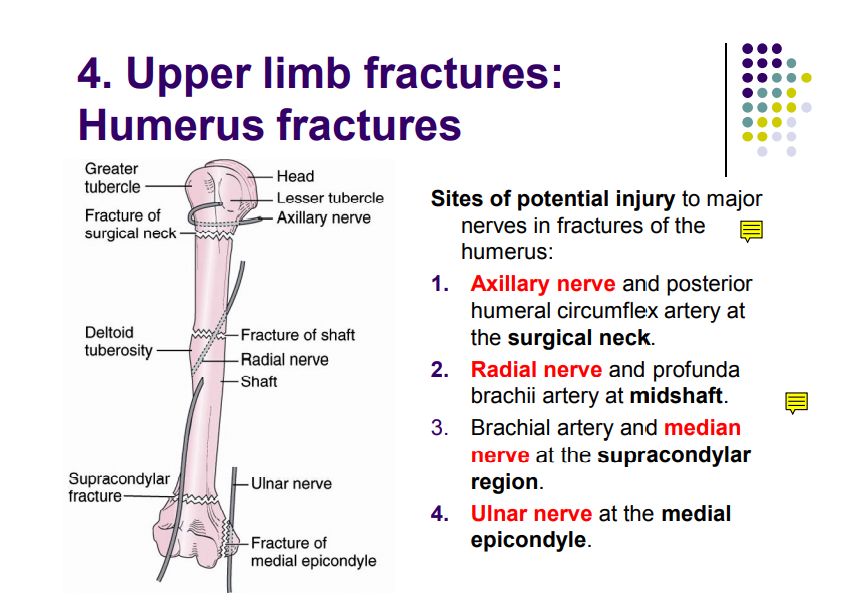
1. Musculocutaneous nerve (lateral cord, C5-7) 2. Median nerve (lateral + medial cords, C5-T1) 3. Ulnar nerve (medial cord, C8-T1) 4. Radial nerve (posterior cord, C5-T1) 5. Axillary nerve (posterior cord, C5-6) 6. Thoracodorsal nerve (posterior cord, C6-8 → latissimus dorsi) — plus long thoracic (C5-7, roots → serratus anterior) & medial/lateral pectoral nerves
- Formed by anterior rami of C5–T1; roots → trunks → divisions → cords → branches
- Roots: C5, C6, C7, C8, T1
- Trunks: upper (C5-6), middle (C7), lower (C8-T1)
- Divisions: each trunk splits into anterior & posterior
- Cords: lateral (anterior divisions of upper & middle), medial (anterior division of lower), posterior (all posterior divisions)
- Six main branches to remember (the "terminal" or clinically key branches):
- Relations to axillary artery: lateral cord = lateral to part 2; medial cord = medial; posterior cord = posterior (hence names)
- Clinical: the plexus is at risk in the axilla during surgery/trauma; Erb's palsy (C5-6, upper trunk), Klumpke's palsy (C8-T1, lower trunk) — see Ch10
7. Axillary lymph nodes (groups)
- Anterior (pectoral) — receive breast & anterior thoracic wall
- Posterior (subscapular) — receive back & posterior shoulder
- Lateral (humeral) — receive upper limb (except areas drained by deltopectoral nodes)
- Central — receive all the above
- Apical — receive central + cephalic route; drain to supraclavicular nodes
- Mnemonic for axillary node order: "Anterior, Posterior, Lateral → Central → Apical"
High-Yield Points
- Axillary artery branches by part (mnemonic): "Screw The Lawyer, Save A Patient" (1st: Superior thoracic; 2nd: Thoracoacromial + Lateral thoracic; 3rd: Subscapular + Anterior & Posterior humeral circumflex)
- Six key plexus branches: musculocutaneous, median, ulnar, radial, axillary, thoracodorsal (long thoracic & pectoral nerves also classic)
- Breast lymphatics: 75% → axillary (anterior/pectoral first); medial → internal thoracic; MCQ answer
- Clavipectoral fascia pierced by: cephalic vein, thoracoacromial vessels, lateral pectoral nerve (classic list)
- Axillary boundaries mnemonic: "A-P-M-L" = Anterior (pec major/minor), Posterior (latissimus/teres major/subscapularis), Medial (serratus anterior), Lateral (bicipital groove)
- Axillary artery injury: compress against humerus; ligation may cause arm ischemia (collaterals: scapular anastomoses)
- Long thoracic nerve injury (during axillary node clearance): winged scapula (serratus anterior paralysis)
Topic Summary
The pectoral region & axilla contain the breast (draining 75% to axillary nodes), the axillary artery (3 parts: Screw-The-Lawyer-Save-A-Patient), the axillary vein, the brachial plexus (C5-T1, six key branches), and the axillary lymph nodes (anterior→central→apical). The clavipectoral fascia is pierced by the cephalic vein, thoracoacromial vessels & lateral pectoral nerve. Injuries during axillary surgery may affect the long thoracic nerve (winged scapula) or medial pectoral nerve (pectoralis major weakness).
LMCHK OSCE Practice
- Breast examination: systematic inspection (dimpling, peau d'orange, nipple) + palpation in quadrants + axillary lymph node palpation (describe anterior/posterior/lateral/central/apical groups).
- Axillary nerve & shoulder abduction: axillary nerve (C5-6) supplies deltoid & teres minor — injury → quadrate shoulder (flattened deltoid), weak abduction.
- Winged scapula: push against a wall → scapula wings off the chest wall (long thoracic nerve → serratus anterior).
- Thoracic outlet test: Adson's maneuver — diminished radial pulse on arm abduction/rotation with cervical rib (compresses subclavian a. & lower trunk of plexus).
- Brachial plexus screen: test each major nerve quickly (shoulder abduction = axillary, elbow flexion = musculocutaneous, wrist flexion = median, spread fingers = ulnar, wrist extension = radial).
Illustrations(图解速览)
Regional anatomy diagrams — identify the structures shown with the chapter content: