# Ch14: Thorax — Wall, Pleura, Lungs & Mediastinum
Preparatory Mindset
The thorax chapter covers the thoracic wall (intercostal spaces), the pleura & pleural cavity, the lungs & their roots, and the mediastinum (boundaries, divisions, contents). The lecture's key points: (1) arrangement of the thoracic wall, (2) the order & course of the internal intercostal arteries/veins/nerves in the costal groove, (3) contents of the mediastinum.
Exam mindset: Intercostal neurovascular bundle order (VAN), the pleura recesses, the lung root relationships (right vs left hilum), and the mediastinum divisions are the classic questions. The Shelf Notes add diaphragm (concept 22), heart (23-25), lung segments (26-27), and pneumothorax (28).
Core Concepts
1. Thoracic wall & intercostal spaces
- Vein, Artery, Nerve — mnemonic "VAN" - Position: runs in the costal groove of the rib above, between the internal & innermost intercostal muscles - Clinical: needle/chest drain should be inserted superior to the rib below (upper border of the lower rib) to avoid the bundle; the bundle is highest at the angle of the rib, more exposed anteriorly
- Layers of the wall: skin → superficial fascia → deep fascia → muscles (pectoralis major/minor, serratus anterior) → ribs & intercostal muscles → endothoracic fascia → parietal pleura
- Intercostal muscles: external (fibers down & forward — like hands in pockets), internal (down & back), innermost
- Intercostal neurovascular bundle (in the costal groove, from above down):
- Internal thoracic (mammary) artery: descends 2 cm lateral to the sternum; gives anterior intercostal branches; used for coronary bypass (LIMA to LAD)
2. Pleura & pleural cavity
- Costodiaphragmatic recess (deepest) — at the costodiaphragmatic angle; fluid collects here (pleural effusion) - Costomediastinal recess
- Parietal pleura (lines the wall) vs visceral pleura (covers the lung); continuous at the hilum (pulmonary ligament hangs below)
- Parietal pleura parts: costal, mediastinal, diaphragmatic, cervical (cupula) — the cupula rises 2-3 cm above the medial 1/3 of the clavicle
- Pleural recesses (where lungs don't fill):
- Nerve supply of pleura: parietal = phrenic nerve (mediastinal & diaphragmatic parts — referred pain to shoulder via C3-5!) & intercostal nerves (costal); visceral = autonomic (pain-insensitive)
- Clinical: pleuritic pain (parietal inflammation); shoulder-tip pain (diaphragmatic pleura → phrenic → C3-5); pneumothorax (air in the pleural cavity — concept 28: open pneumothorax = sucking chest wound → mediastinal flutter → urgent seal); hemothorax, effusion, empyema
3. Lungs & their roots (hila)
- Right: "BAPA" — Bronchus (upper, posterior), Artery (pulmonary, anterior), Pulmonary veins (inferior) — from above: bronchus, artery, veins (the bronchus is posterior & superior; the eparterial bronchus on the right: the upper lobe bronchus arises ABOVE the pulmonary artery) - Left: "BAPA" reversed — Artery (upper), Bronchus, Pulmonary veins (inferior) — the bronchus is inferior to the artery (hyparterial); left main bronchus is longer, narrower & more horizontal than the right (right main bronchus is shorter, wider, more vertical → aspiration goes to the right)
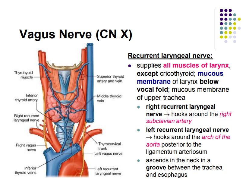
- Right side: middle = root; superior = azygos arch; anteroinferior = pericardium; posterior = esophagus, azygos vein, sympathetic trunk, right vagus; anterior = right phrenic n. + pericardiacophrenic vessels - Left side: middle = root; superior = aortic arch; anteroinferior = pericardium; posterior = esophagus, thoracic aorta, sympathetic trunk, left vagus; anterior = left phrenic n. + pericardiacophrenic vessels
- Right lung: 3 lobes (superior, middle, inferior), horizontal + oblique fissures; bronchopulmonary segments: upper 3, middle 2, lower 5 (10 segments)
- Left lung: 2 lobes (superior, inferior), oblique fissure only + cardiac notch & lingula; upper 4 + lower 5 = 9 segments (apicoposterior & anteromedial basal fused)
- Mediastinal surface impressions: right = cardiac impression, grooves for SVC, azygos arch, esophagus; left = cardiac impression, grooves for aortic arch & descending aorta, esophagus
- Hilum (root of lung) contents & arrangement (MUST KNOW):
- Relations around the root of the lung (lecture MUST KNOW):
- Clinical: bronchial carcinoma at the hilum; hilar lymphadenopathy; lung cancer invading the phrenic (diaphragm paralysis) or recurrent laryngeal (hoarseness)
4. Mediastinum
- Definition: the region between the two pleural cavities
- Boundaries: superior = superior thoracic aperture; inferior = diaphragm; anterior = sternum; posterior = bodies of T1-T12; lateral = mediastinal parietal pleura
- Divisions: superior (above the sternal angle plane) & inferior (below; split by the pericardium into anterior, middle, posterior mediastina)
- Superior mediastinum contents (structures passing between thorax & neck/upper limb): thymus, brachiocephalic veins & SVC (upper part), arch of aorta (beginning & end at the sternal angle) with its 3 branches, trachea, esophagus, thoracic duct, phrenic & vagus nerves, sympathetic trunks
- Arch of aorta branches (anterior→posterior): brachiocephalic trunk, left common carotid, left subclavian — mnemonic "B-C-S" (Beer Can Stash / Bread Comes Slice)
- Middle mediastinum = pericardium & heart, great vessels (ascending aorta, pulmonary trunk, SVC, IVC)
- Anterior mediastinum: thymus (remnant in adult), lymph nodes, fat
- Posterior mediastinum: esophagus, descending thoracic aorta, azygos & hemiazygos veins, thoracic duct, sympathetic trunks, vagus nerves
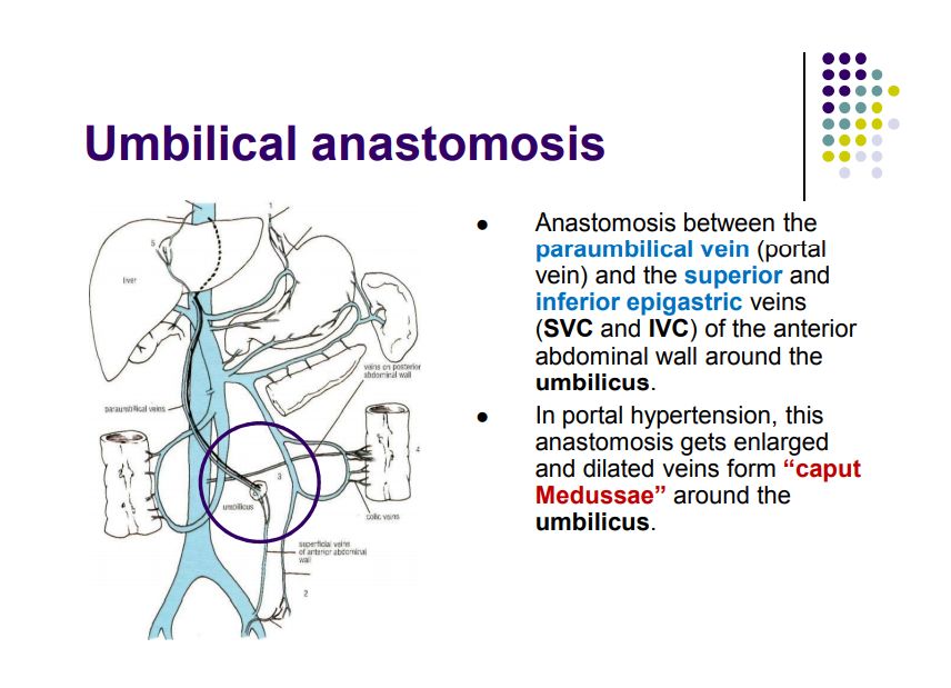
- Clinical: mediastinal masses (thymoma, lymphoma, teratoma, neurogenic); esophageal varices in portal HTN drain to azygos
5. Heart — key relationships (Shelf concepts 23-25)
- Right coronary artery: SA nodal branch, right marginal, posterior interventricular (in 80-85%) — supplies right ventricle & inferior wall; occlusion → inferior MI (usually RCA) - Left coronary artery: left anterior descending (anterior interventricular — "widow maker") & circumflex branches; LAD supplies anterior wall & septum - Dominance: right dominant in most people
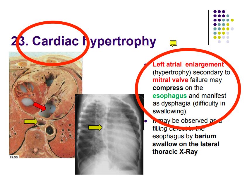
- Cardiac hypertrophy & chamber enlargement change the cardiac silhouette on CXR (see Imaging notes)
- Auscultation areas (concept 24): aortic = 2nd right intercostal space; pulmonary = 2nd left ICS; tricuspid = 4th/5th left ICS near sternum (lower left sternal border); mitral = 5th left ICS midclavicular line (apex)
- Blood supply (concept 25):
6. Diaphragm (concept 22)
- T8: IVC (+ right phrenic nerve) - T10: esophagus + vagus nerves (esophageal hiatus) - T12: aorta + thoracic duct (+ azygos vein) - Mnemonic: "I (8) ate (10) at (12)" — IVC at T8, Esophagus at T10, Aorta at T12
- Structure: muscular dome; central tendon; separates thorax from abdomen
- Openings (levels):
- Nerve supply: phrenic nerve (C3-C5), motor only; sensory = phrenic (central) + lower intercostals (peripheral)
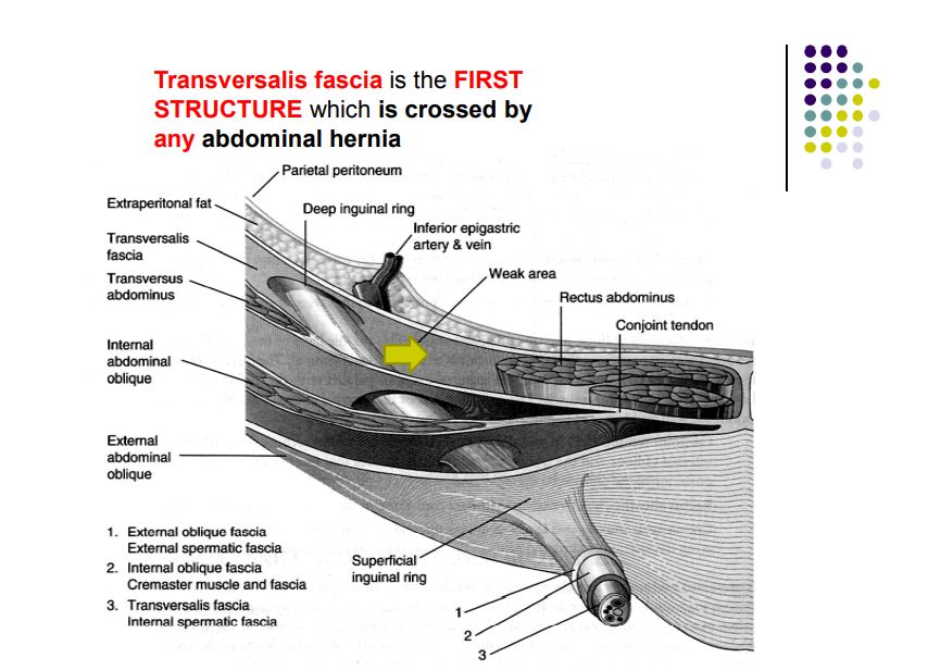
- Clinical: congenital diaphragmatic hernia (Bochdalek — posterolateral, more common left; concept 33); sliding hiatal hernia (concept 34: stomach & GE junction above diaphragm → reflux); paralysis → paradoxical movement (sniff test)
High-Yield Points
- Intercostal bundle: "VAN" in the costal groove (vein uppermost, nerve lowest); insert chest drains/needles on the upper border of the rib
- Pleural recesses: costodiaphragmatic = deepest — fluid collects here
- Right hilum "BAPA" (bronchus upper & posterior, artery anterior, veins inferior); left hilum artery upper — plus right main bronchus more vertical → aspiration to right
- Root of lung relations: phrenic nerve & pericardiacophrenic vessels anterior to the root on both sides; vagus posterior
- Mediastinum: superior (above sternal angle) + anterior/middle/posterior inferior
- Arch branches (anterior→posterior): brachiocephalic trunk, LCCA, LSA ("BCS")
- Diaphragm openings: "I ate at 12" — IVC T8, Esophagus T10, Aorta T12
- Auscultation: aortic 2R, pulmonary 2L, tricuspid lower left sternal border, mitral apex
- Coronary arteries: RCA → inferior (posterior IV in most); LAD → anterior wall & septum ("widow maker"); circumflex → lateral
- Aspiration goes to the right lower lobe (more vertical bronchus)
- Pneumothorax (concept 28): open = sucking wound → seal it; tension = mediastinal shift, hyperresonance, absent breath sounds → needle decompression
Topic Summary
The thoracic wall carries the intercostal neurovascular bundle (VAN) in the costal groove; pleural recesses (costodiaphragmatic) are where fluid accumulates. The lungs have 3 (right) / 2 (left) lobes; at the hilum the right bronchus sits above the artery (BAPA) while the left artery sits above the bronchus; the right main bronchus is more vertical (aspiration site). The mediastinum is divided into superior and three inferior parts (anterior/middle/posterior); its key contents include the arch of aorta (BCS branches), thymus, trachea, esophagus, thoracic duct, and nerves. The diaphragm (phrenic C3-C5) has openings at T8 (IVC), T10 (esophagus), T12 (aorta). The heart's blood supply (RCA/LAD/circumflex) and auscultation areas complete the picture.
LMCHK OSCE Practice
- Chest drain insertion: describe the safe triangle (anterior to midaxillary line, 4th-5th ICS, above the rib) — explain why (intercostal VAN bundle in the costal groove).
- Chest examination: percussion & auscultation; dullness = effusion/consolidation, hyperresonance = pneumothorax; describe the costodiaphragmatic recess.
- Cardiac auscultation: demonstrate the four valve areas; explain where murmurs radiate (aortic → carotids; mitral → axilla).
- Tension pneumothorax emergency: tracheal deviation away, hyperresonance, absent breath sounds → immediate needle decompression (2nd ICS midclavicular line).
- Diaphragm assessment: sniff test on fluoroscopy; describe paradoxical movement with phrenic palsy.
- Aspiration scenario: an elderly patient aspirates while supine → right lower lobe pneumonia — explain the bronchial anatomy (right main bronchus more vertical & wider).
Illustrations(图解速览)
Regional anatomy diagrams — identify the structures shown with the chapter content: