# Ch19: Pelvis & Perineum
Preparatory Mindset
The pelvis is divided into the greater (false) & lesser (true) pelvis by the terminal line (pelvic brim). The lecture's key points: (1) composition & division of the pelvis & pelvic diaphragm, (2) positions & relationships of pelvic organs, (3) course, branches & distribution of the internal iliac artery, (4) boundaries & contents of the ischiorectal fossa, (5) superficial & deep perineal spaces. Male vs female pelvic organ arrangement is a classic table question.
Exam mindset: Internal iliac artery branches (parietal vs visceral), the pelvic diaphragm & its hiatus, the ischiorectal fossa, and the male/female organ differences are the core. The 100-Concepts deck adds perineal pouches (54-55), male urethra (61), prostate (60), uterus (66-67), lymphatics (64-65), and hemorrhoids (53).
Core Concepts
1. Bony pelvis
- Greater (false) pelvis: above the pelvic inlet — contains the lower abdominal viscera (cecum, sigmoid colon, coils of ileum) - Lesser (true) pelvis: between the pelvic inlet & outlet — contains the pelvic viscera (bladder, rectum, reproductive organs)
- Female: wider, shallower; pubic arch 90-100°; oval inlet; larger pelvic outlet (for childbirth) - Male: narrower, deeper; pubic arch 70-75°; heart-shaped inlet
- Composition: 2 hip bones + sacrum + coccyx
- Division (by the terminal line / pelvic brim):
- Pelvic inlet (terminal line/brim): sacral promontory, arcuate line, pecten pubis, pubic tubercle, pubic crest, superior margin of pubic symphysis
- Pelvic outlet: inferior margin of pubic symphysis, ischiopubic ramus, ischial tuberosity, sacrotuberous ligament, tip of coccyx
- Male vs female pelvis:
- Landmarks: iliac crest, ASIS, PSIS, pubic tubercle, pubic symphysis/crest, tubercle of iliac crest, ischial tuberosity
2. Pelvic wall & pelvic floor
- Levator ani (main): puborectalis, pubococcygeus, iliococcygeus (+ levator prostatae/pubovaginalis in males/females) - Coccygeus (ischiococcygeus) - Tendinous arch of levator ani (fascial origin)
- Lateral wall: obturator internus muscle
- Posterior wall: piriformis muscle
- Pelvic floor (pelvic diaphragm):
- Hiatus of the pelvic diaphragm: anterior gap, closed by the urogenital diaphragm; perforated by the urethra (male) and urethra + vagina (female); the rectum passes through the posterior part (levator ani loop — puborectalis forms the anorectal angle)
- Clinical: pelvic floor weakness → cystocele (concept 56), rectocele, uterine prolapse; Kegel exercises strengthen levator ani; the pelvic diaphragm supports the pelvic viscera & maintains continence
3. Pelvic fascia & spaces
- Parietal pelvic fascia: lines the walls & muscles
- Visceral pelvic fascia: covers organs, forms septa: rectovesical septum (male), rectovaginal & vesicovaginal septa (female)
- Spaces: retropubic space (between pubic symphysis & bladder — site of retropubic prostatectomy/space of Retzius), pararectal space, retrorectal space (between rectal fascia & presacral fascia)
4. Internal iliac artery (MUST KNOW)
- Iliolumbar artery - Lateral sacral arteries - Superior gluteal artery (through the greater sciatic foramen above piriformis) - Inferior gluteal artery (through the greater sciatic foramen below piriformis) - Obturator artery (through the obturator canal) — mnemonic "I L S S O" (Iliolumbar, Lateral sacral, Superior gluteal, ... Obturator)
- Umbilical artery → superior vesical artery - Inferior vesical artery (male: also to prostate) - Uterine artery (female) - Vaginal artery (female; also inferior vesical in male) - Middle rectal artery - Internal pudendal artery (leaves the pelvis through the infrapiriform foramen, re-enters via the lesser sciatic foramen with the pudendal nerve — supplies the perineum)
- Origin: common iliac artery bifurcation at L5/S1 (in front of the sacroiliac joint)
- Divisions: anterior (visceral) & posterior (parietal) trunks
- Parietal branches (posterior trunk):
- Visceral branches (anterior trunk):
- Mnemonic for visceral: "U U V M I P" (Umbilical, Uterine, Vesical, Middle rectal, Internal pudendal)" — better: "Uterine, Vesical, Middle rectal, Internal pudendal"
- Uterine artery (MUST KNOW): crosses the ureter about 2 cm from the cervix, IN FRONT of it ("water under the bridge" — the ureter passes under the uterine artery) — ligate the uterine artery in hysterectomy while protecting the ureter
- Internal iliac vein: tributaries = rectal, vesical, vaginal, prostatic venous plexuses
5. Lymphatic drainage of the pelvis
- Internal iliac nodes: pelvic viscera, perineum, buttock, back of thigh
- External iliac nodes: lower limb + parts of pelvic viscera
- Sacral nodes: posterior pelvic wall
- Common iliac nodes: receive all → lumbar (para-aortic) nodes
- Clinical: cervical/prostate cancer spread follows these chains
6. Nerves of the pelvis
- Superior hypogastric plexus: in front of L5 between the common iliac arteries - Inferior hypogastric (pelvic) plexus: on each side of the rectum - Sacral sympathetic trunk: 4-5 ganglia
- Sacral plexus (L4-S4): formed by the lumbosacral trunk (L4-5) + anterior rami of S1-S4; lies anterior to piriformis; branches: sciatic, pudendal, superior/inferior gluteal, posterior femoral cutaneous
- Autonomic plexuses:
7. Pelvic viscera — male vs female (MUST KNOW)
| Region | Male | Female |
|---|---|---|
| Anterior | Urinary bladder, prostatic urethra, prostate | Urinary bladder, urethra |
| Middle | Ductus deferens, seminal vesicle, ejaculatory duct | Uterus, vagina, uterine tubes, ovaries |
| Posterior | Rectum | Rectum |
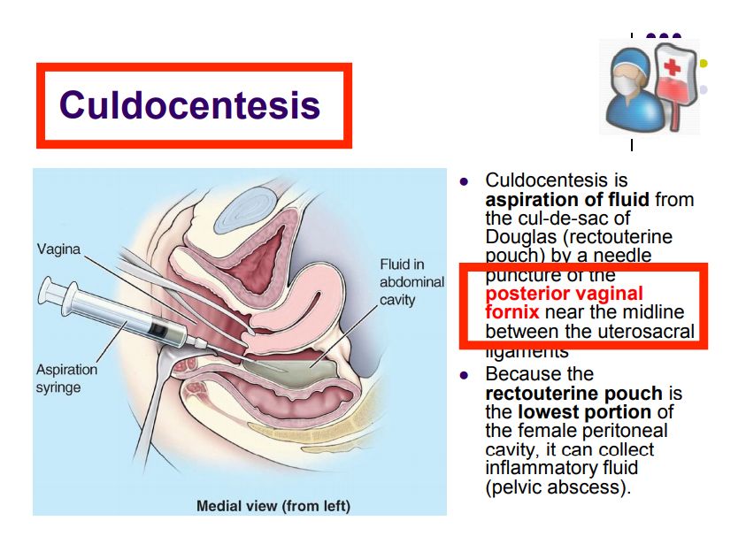
- Urinary bladder: behind the pubic symphysis (retropubic space); covered superiorly by peritoneum; suprapubic (extraperitoneal) aspiration/catheterization avoids the peritoneum (concept 59: paracentesis of the bladder — the needle enters above the pubic symphysis, piercing the anterior wall without peritoneum)
- Prostate (concept 60): surrounds the prostatic urethra; palpated by digital rectal exam (DRE); zones: peripheral (70% of cancers — palpable), central, transition (BPH); lymphatic drainage → internal iliac/obturator nodes; "prostate tumors" spread to pelvic lymph nodes & bone (lumbar spine)
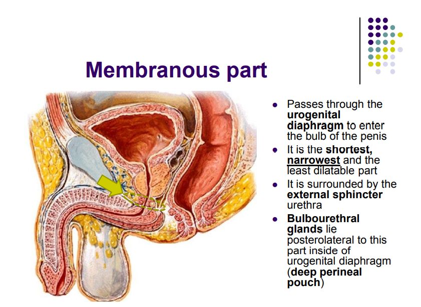
- Male urethra (concept 61 — MUST KNOW): 4 parts: preprostatic (intramural), prostatic (widest & most dilatable; urethral crest, seminal colliculus), membranous (narrowest, through the urogenital diaphragm, site of trauma), spongy (penile, longest; bulbous & penile portions) — mnemonic "P M S" (Prostatic, Membranous, Spongy). Rupture of the membranous urethra (straddle injury/pelvic fracture) → urine extravasates into the superficial perineal pouch (scrotum, penis, anterior abdominal wall via Scarpa's)
- Rectum & anal canal: hemorrhoids (concept 53): internal (above the pectinate line — from superior rectal artery, painless bright-red bleeding, covered by mucous membrane) vs external (below the pectinate line — from inferior rectal artery, painful, covered by skin); the pectinate line is the embryological watershed (superior rectal = portal; inferior rectal = systemic)
- Female organs (concepts 66-67): uterus (fundus, body, cervix; anteverted/anteflexed); arterial supply = uterine artery (from internal iliac) + ovarian artery (from aorta); parts of the uterine tube (concept 67): infundibulum (with fimbriae), ampulla (fertilization site), isthmus, intramural/uterine part; ectopic pregnancy most often in the ampulla
8. Perineum — ischiorectal fossa & perineal pouches (concepts 54-55)
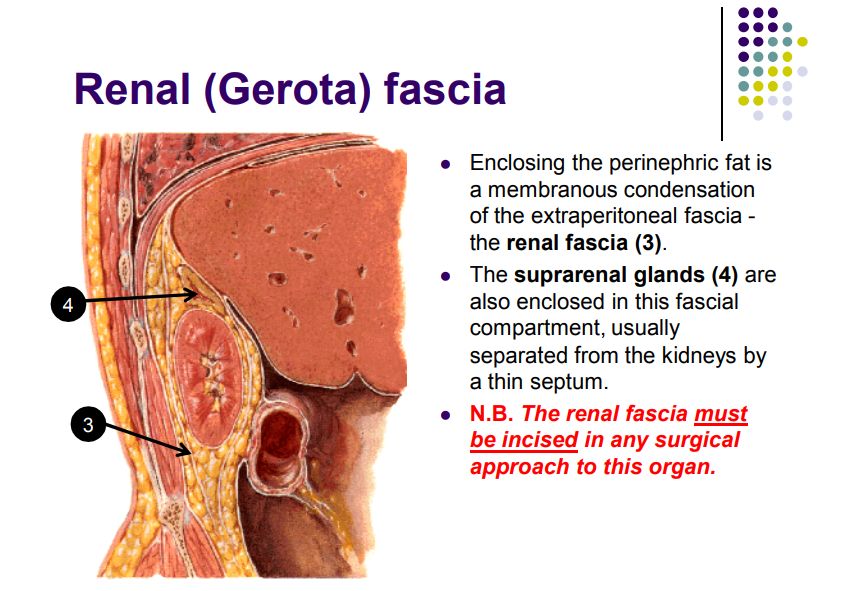
- Ischiorectal (ischioanal) fossa: wedge-shaped space on each side of the anal canal; boundaries: medial = levator ani & external anal sphincter; lateral = obturator internus fascia & ischial tuberosity; posterior = sacrotuberous ligament & gluteus maximus; anterior = urogenital diaphragm; contents: fat, inferior rectal vessels & nerves, pudendal canal (Alcock's) in the lateral wall with the internal pudendal vessels & pudendal nerve
- Clinical: ischiorectal abscess → fistula-in-ano; pudendal nerve block (through the ischiorectal fossa toward the ischial spine)
- Deep perineal pouch (concept 54): within the urogenital diaphragm; contents: sphincter urethrae, deep transverse perineal muscle, bulbourethral (Cowper's) glands (male only) — ducts perforate the perineal membrane
- Superficial perineal pouch (concept 55): between the perineal membrane & Colles' fascia; contents: ischiocavernosus, bulbospongiosus, superficial transverse perineal muscles; crura of the penis/clitoris, bulb of the penis, greater vestibular (Bartholin's) glands (female), branches of the internal pudendal vessels & pudendal nerve
- Clinical: Bartholin cyst (infection of the greater vestibular gland); superficial perineal pouch is the site of urine extravasation in membranous urethral rupture
High-Yield Points
- Internal iliac artery: bifurcation at L5/S1; posterior trunk = iliolumbar, lateral sacral, superior gluteal, inferior gluteal, obturator; anterior trunk = umbilical (→ superior vesical), inferior vesical, uterine, vaginal, middle rectal, internal pudendal
- Uterine artery crosses the ureter ~2 cm from the cervix (in front of the ureter — "water under the bridge")
- Internal pudendal artery + pudendal nerve: greater sciatic (infrapiriform) → lesser sciatic → pudendal canal → perineum
- Pelvic diaphragm: levator ani (puborectalis, pubococcygeus, iliococcygeus) + coccygeus; hiatus closed by the urogenital diaphragm (urethra; urethra+vagina)
- Male urethra: prostatic (widest) → membranous (narrowest, trauma) → spongy (longest) — membranous rupture → superficial pouch extravasation
- Pectinate line: internal hemorrhoids above (superior rectal, portal); external below (inferior rectal, systemic)
- Uterine tube parts: infundibulum, ampulla (fertilization), isthmus, intramural
- Ischiorectal fossa: pudendal canal (Alcock's) in the lateral wall; fat, inferior rectal n./v.
- Superficial perineal pouch: crura/bulb, ischiocavernosus, bulbospongiosus, superficial transverse perineal, Bartholin's glands (female)
- Deep perineal pouch: sphincter urethrae, deep transverse perineal, Cowper's glands (male)
- Prostate: peripheral zone = cancer; transition = BPH; DRE
- Bladder: suprapubic access is extraperitoneal (above the pubic symphysis)
- Female pelvis: pubic arch 90-100° (male 70-75°)
Topic Summary
The pelvis (greater vs lesser by the terminal line) is floored by the pelvic diaphragm (levator ani + coccygeus; hiatus for urethra ± vagina) and supplied by the internal iliac artery (posterior trunk: iliolumbar, lateral sacral, superior/inferior gluteal, obturator; anterior trunk: umbilical→superior vesical, inferior vesical, uterine, vaginal, middle rectal, internal pudendal). The uterine artery crosses in front of the ureter — the key surgical relation. Male/female organs differ in the middle layer (ductus deferens/seminal vesicle vs uterus/tubes/ovaries). The male urethra has prostatic (widest), membranous (narrowest, trauma) & spongy parts; the ischiorectal fossa (pudendal canal) and the superficial/deep perineal pouches (Bartholin's/Cowper's glands) complete the perineum. Hemorrhoids above/below the pectinate line reflect portal vs systemic drainage.
LMCHK OSCE Practice
- Digital rectal examination (DRE): palpate the prostate (size, nodule, tenderness), the cervix (female), and the rectum; describe the ischiorectal fossa relations.
- Male catheterization: describe the urethral anatomy (3 parts, 2 curves) and why prostatic enlargement (BPH) obstructs catheterization.
- Urinary retention / suprapubic catheter: describe the extraperitoneal suprapubic approach (bladder behind the pubic symphysis).
- Pelvic examination (female): describe the uterus position & the uterine artery/ureter relation (risk in hysterectomy).
- Hemorrhoid exam: differentiate internal (above pectinate line, painless bleeding) from external (below, painful thrombosis); describe the venous drainage (superior rectal → portal vs inferior rectal → systemic).
- Perineal trauma (straddle injury): membranous urethral rupture → blood at the meatus, urine extravasation into the superficial pouch — describe the fascial planes (Colles'/Scarpa's → scrotum, penis, anterior abdominal wall).
- Pudendal nerve block: describe the approach via the ischiorectal fossa toward the ischial spine (for forceps delivery/episiotomy).
- Sacral plexus screen: foot plantarflexion (tibial), dorsiflexion (deep peroneal), ankle jerk (S1) — same reflexes as the lower limb chapter.
Illustrations(图解速览)
Regional anatomy diagrams — identify the structures shown with the chapter content: