# Ch02: Lower Limb I — Superficial Structures & Surface Anatomy
Preparatory Mindset
The lower limb is divided into four parts: gluteal region, thigh, leg, foot. This chapter covers the superficial structures — the layer between skin and deep fascia — because it is where clinicians cannulate veins, feel lymph nodes, and find cutaneous nerves. The single most important structure is the great saphenous vein.
Exam mindset: "Where does the great saphenous vein run? What are its tributaries? Where do the superficial lymph nodes lie?" are guaranteed questions. Know the surface landmarks perfectly (ASIS, pubic tubercle, inguinal ligament, patella, medial/lateral femoral epicondyles, head & neck of fibula, medial/lateral malleoli).
Core Concepts
1. Surface landmarks & division of the lower limb
- Landmarks: iliac crest, ASIS, pubic tubercle, inguinal ligament, patella, medial & lateral femoral epicondyles, head & neck of fibula, medial & lateral malleoli
- Division: gluteal region / thigh / leg / foot
2. Superficial structures of lower limb
- Skin
- Superficial fascia contains: superficial arteries & veins, lymphatic vessels & nodes, cutaneous nerves
3. Great saphenous vein (MUST KNOW — "must to know" in lecture)
- Origin: medial end of the dorsal venous arch of the foot
- Course: passes anterior to the medial malleolus (key landmark!) → ascends along the medial side of the leg → passes posterior to the medial condyle of the femur → ascends the medial thigh → pierces the saphenous opening (fascia lata hiatus) → drains into the femoral vein
- Tributaries (near the saphenous opening): superficial external pudendal, superficial epigastric, superficial circumflex iliac veins; plus (lower) the accessory saphenous veins
- Clinical: great saphenous vein is used for coronary artery bypass grafting (CABG) and for long saphenous cutdown; varicose veins arise here; it is at risk during medial malleolus surgery
4. Saphenous nerve & small saphenous vein
- Saphenous nerve (from femoral nerve): accompanies the great saphenous vein in the leg, where it lies anterior (lateral) to the vein — know this relation (lecture: "Relation of the saphenous nerve and greater saphenous vein in the leg")
- Small saphenous vein: arises from the lateral end of the dorsal venous arch, ascends posterior to the lateral malleolus, runs in the posterior leg, pierces deep fascia and drains into the popliteal vein in the popliteal fossa
5. Superficial lymph nodes of lower limb
- Superficial inguinal nodes (upper group along the inguinal ligament; lower group near the saphenous opening) — drain most of the lower limb
- Deep inguinal nodes — receive deep lymphatics + superficial nodes, drain to external iliac nodes
- Popliteal nodes — in the popliteal fossa, drain the leg & foot
- Clinical: palpable inguinal nodes with lower-limb infection; sentinel node in melanoma
6. Superficial branches of the femoral artery
- Superficial epigastric, superficial circumflex iliac, superficial external pudendal arteries — all supply the skin/fascia of the lower abdomen & groin, arise from the femoral artery near the inguinal ligament
- Deep external pudendal artery also from femoral
High-Yield Points
- Mnemonic for the great saphenous vein course: "Medial malleolus → medial knee → saphenous opening → femoral vein" — it NEVER crosses the lateral side
- Key relation: great saphenous vein lies anterior to medial malleolus; small saphenous vein lies posterior to lateral malleolus
- Saphenous nerve is at risk when stripping the great saphenous vein (causes medial leg numbness)
- CABG: great saphenous vein is the classic conduit; also used for dialysis access
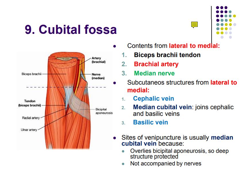
- Venipuncture: the dorsal venous arch / great saphenous at the ankle is a common IV site
- Lymph drainage: superficial inguinal nodes → deep inguinal nodes → external iliac → common iliac → lumbar nodes
Topic Summary
The superficial layer of the lower limb contains the great saphenous vein (the key vessel — anterior to medial malleolus, ascending to the saphenous opening, draining into the femoral vein), the small saphenous vein (posterior to lateral malleolus → popliteal vein), superficial branches of the femoral artery, the saphenous nerve (femoral n., anterior to the great saphenous vein in the leg), and the inguinal/popliteal lymph node groups. Surface landmarks anchor every clinical procedure from IV access to hernia surgery.
LMCHK OSCE Practice
- Examine the leg for varicose veins: describe the course of the great saphenous vein and its tributaries; know that incompetence of valves causes reflux.
- Cannulation site: great saphenous vein at the ankle (anterior to medial malleolus) or dorsal venous arch — be ready to say why (superficial, accessible, no artery immediately adjacent at the ankle).
- Palpate inguinal lymph nodes: horizontal (upper) and vertical (lower) groups — describe drainage of the lower limb to the examiner.
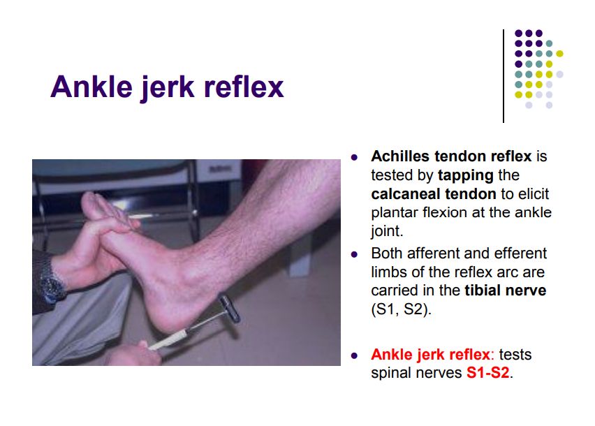
- Tendon reflexes: the knee jerk (L3/L4) and ankle jerk (S1/S2) rely on the same segmental levels you will be asked about in a neuro exam.
Illustrations(图解速览)
Regional anatomy diagrams — identify the structures shown with the chapter content: