Preparatory Mindset
Epidemic Haemorrhagic Fever (EHF) — also called Haemorrhagic Fever with Renal Syndrome (HFRS) since the WHO renaming in 1982 — is a China-endemic rodent-borne Hantavirus infection with a classic 5-phase clinical course: fever → shock → oliguria → diuresis → convalescence (CM exam tested). The exam mindset: triad of fever + haemorrhage + renal failure in a patient with rural/forest exposure → think EHF. The kidneys tell the story — proteinuria + atypical lymphocytes + rising BUN/Cr appear as early as day 2. Compared with dengue, EHF adds renal failure and shock; compared with sepsis, EHF has a biphasic course and renal dominance. Recognition in the febrile phase saves lives — primary shock accounts for 33% of deaths, oliguria-phase complications for 50%.
Core Concepts
1. Definition
A natural-source infectious disease caused by Hantaviruses (family Bunyaviridae, now Hantaviridae), characterised by the triad of fever, haemorrhage, and renal failure, classically passing through 5 progressive phases.
2. Aetiology — Hantavirus
| Feature | Detail |
|---|---|
| Family | *Bunyaviridae* (Hantaviridae genus) |
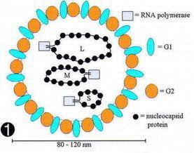
| Genome | Single-strand, negative-sense RNA; 3 segments — L (polymerase), M (envelope glycoproteins G1, G2 — G2 contains the neutralising antigen, vaccine target), S (nucleocapsid protein — strong immunogenicity, complement-fixing antigen) |
| Morphology | Spherical or oval, 80–120 nm diameter, envelope with surface projections |
| Stability | Stable up to 2 weeks; inactivated by pH <5, ethanol, ether, chloroform, 60 °C/30 min, 100 °C/1 min, UV |
| Important species | Hantaan (HTNV) — severe HFRS; Seoul (SEOV) — moderate HFRS; Puumala (PUUV) — mild (Europe); Dobrava-Belgrade (DEOV) — severe HFRS; New World (Sin Nombre, Andes) → Hantavirus Cardiopulmonary Syndrome (HCPS/HPS), NOT HFRS |
In China, the epidemic serotypes are Hantaan and Seoul viruses → HFRS is more severe than in Europe.
3. Epidemiology
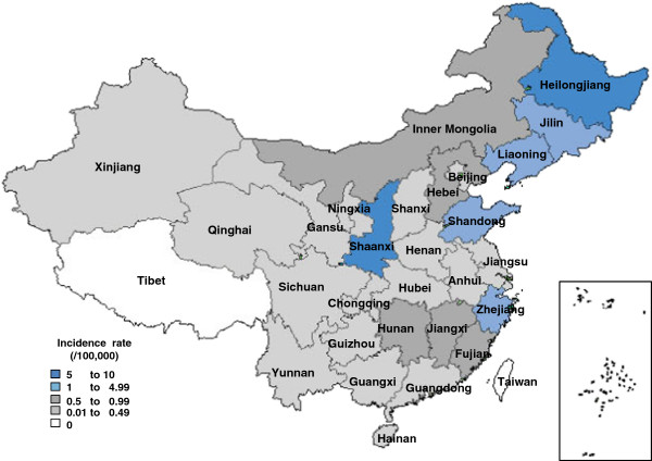
- Distribution: endemic in a belt from Norway → Sweden → Finland → Russia → China → Korea → Japan. China is one of the most seriously affected countries.
- China case load: 50,000–100,000 cases/year from 1980–2000; fell to 20,000–40,000/year after 2001 with vaccination and rodent control.
- Age/sex: predominantly young adult males (rural/forest exposure).
4. Three links of the infectious chain
1. Respiratory (aerosolised rodent excreta — most common) 2. Contact (broken skin/mucosa with contaminated materials) 3. Digestive (contaminated food/water) 4. Vertical (rare; transplacental)
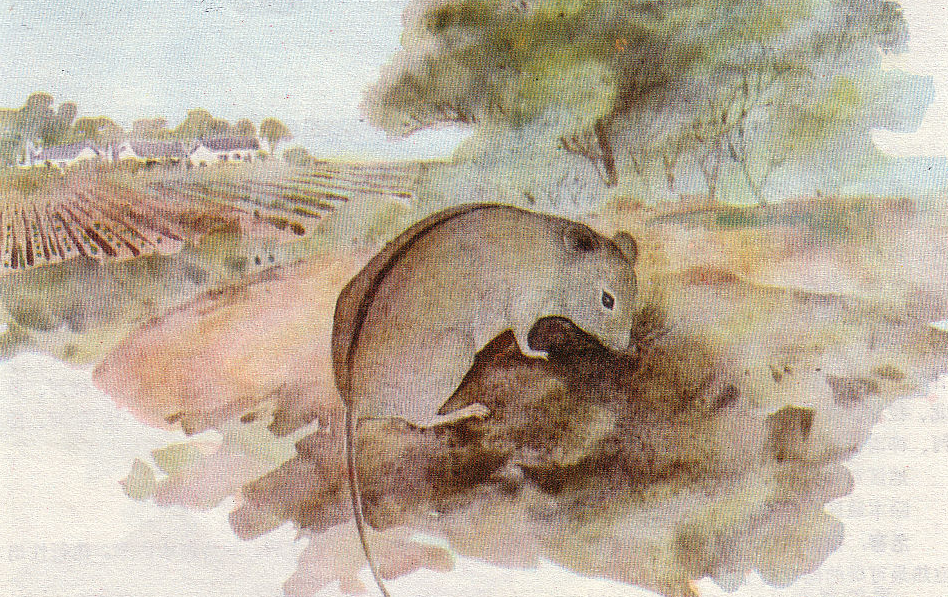
- Source of infection: >170 animal species carry Hantavirus. Rodents are the main reservoir — *Apodemus agrarius* (striped field mouse, 黑线姬鼠), *Rattus norvegicus* (Norway rat, 褐家鼠), *Apodemus peninsulae* (大林姬鼠, forest areas). Also dogs, cats, rabbits. Patients are NOT important sources.
- Route of transmission:
- Susceptible population: non-immune individuals; no gender, age, or occupation bias other than rural exposure.
5. Pathogenesis — six mechanisms of organ damage
- Direct viral injury to small-vessel endothelium (capillary leak, plasma exudation)
- Immune complex deposition → small-vessel and renal tubule damage
- Renal interstitial haemorrhage and oedema → compresses renal tubules
- Renal tissue necrosis (medulla) → pathognomonic lesion
- Activation of renin–angiotensin → afferent arteriolar constriction → ↓ GFR
- Tubular obstruction by protein casts and necrotic debris
Three downstream syndromes follow:
| Syndrome | Mechanism |
|---|---|
| Shock | Plasma exudation → hypovolaemia; secondary shock after oliguria from insufficient fluid replacement |
| Haemorrhage tendency | Vessel wall damage, thrombocytopenia + dysfunction, uraemic platelet defect, heparin-like substances, DIC |
| Acute renal failure | Sum of mechanisms 1–6 above |
6. Pathology
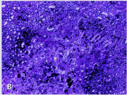
Most affected organ: small vessels and kidneys. Pathognomonic lesion in renal medulla — well-defined necrotic lesion surrounded by haemorrhage and congestion. Similar small-vessel injury in heart, liver, brain.
7. Clinical manifestations
Incubation: 4–46 days, usually 1–2 weeks.
Classic five-phase course (overlapping or skip possible, especially mild/very-severe types):
| Phase | Duration | Hallmarks |
|---|---|---|
| 1. Febrile phase | 3–7 days (≤10) | Acute-onset fever 39–40 °C; "three ache" (headache, eye pain, lumbar pain — small-vessel dilation / oedema); GI symptoms (nausea, vomiting, abdominal pain, diarrhoea); "three flush" — face, neck, chest skin erythema ("drunken" facies); bulbar conjunctival oedema + palpebral/facial oedema; petechiae/ecchymoses on chest, back, axillae; proteinuria appears by day 2 — sometimes with casts, RBCs, and "membrane-like substance" |
| 2. Hypotensive phase | hours to days | BP fall, hypovolaemic shock (primary shock — 33% of deaths); worsening bleeding (petechiae, epistaxis, GI, intracranial); ↑ BUN/Cr, ↑ atypical lymphocytes (>10%); leukocytosis, thrombocytopenia |
| 3. Oliguric phase | 3–7 days | Urine <400 mL/d (oliguria), <50 mL/d (anuria); 50% of deaths here from uraemia, pulmonary oedema, cerebral haemorrhage, secondary infection, cardiac failure; severe electrolyte/acid-base disturbance (hyperkalaemia); hypertension from sodium/water retention |
| 4. Diuretic phase | days to weeks | Urine 3–6 L/d (hyposthenuria); three sub-stages — migratory (400–2000 mL/d, BUN/Cr still ↑), early polyuric (≥2000 mL/d, still severe), late polyuric (≥3000 mL/d, recovery); risk of secondary shock from dehydration/electrolyte loss |
| 5. Convalescent phase | 1–3 months | Urine returns to 1000–2000 mL/24 h, appetite returns, recovery; rare complications — neurological sequelae, hypopituitarism, chronic renal failure |
8. Clinical classification
| Type | Criteria |
|---|---|
| Mild | T < 39 °C, mild intoxication, no oliguria, no shock |
| Moderate | T ≥ 39 °C, severe intoxication, drunken facies, conjunctival oedema, haemorrhage, hypotension, oliguria, marked proteinuria |
| Severe | T ≥ 40 °C, severe intoxication, shock, bleeding, oliguria < 5 d or anuria < 2 d |
| Very severe (危重型) | Severe type + any one of: refractory shock, organ bleeding, acute renal failure, cardiac failure/pulmonary oedema, CNS complications, severe secondary infection |
| Atypical | T < 38 °C, atypical symptoms — may be missed |
9. Laboratory examination
| Test | Pattern |
|---|---|
| CBC | Leukocytosis 15–50 × 10⁹/L; neutrophils early, lymphocytes late; atypical lymphocytes 10–15%; ↑ haematocrit/haemoglobin (haemoconcentration); thrombocytopenia |
| Urinalysis | Proteinuria from day 2; casts, RBCs, membrane-like substance |
| Biochemistry | ↑ BUN, ↑ Cr; hyperkalaemia (oliguria), hypokalaemia (diuresis); elevated LDH |
| Coagulation | PT/aPTT prolonged in severe cases; DIC screen |
| Specific diagnosis | Hantavirus IgM (early), IgG (convalescent); RT-PCR for viral RNA; immunohistochemistry on biopsy (research) |
> Virus-triggered leukocytosis is uncommon — the classic viral causes of leukocytosis are HFRS, infectious mononucleosis, Japanese encephalitis, and rabies.
10. Treatment
No specific antiviral. Management is supportive and phase-driven:
| Phase | Key management |
|---|---|
| Febrile | Rest, fluids, antipyretics (avoid aspirin → bleeding); monitor BP, urine, platelets |
| Hypotensive / shock | Aggressive fluid resuscitation (crystalloid, colloid); vasoactive support if refractory; correct acidosis; treat DIC (FFP, platelets) |
| Oliguric | Strict fluid balance (input = output + insensible); diuretics (furosemide) for established oliguria; renal replacement therapy (haemodialysis) for uraemia, hyperkalaemia, fluid overload; avoid nephrotoxic drugs |
| Diuretic | Replace fluid + electrolytes (Na⁺, K⁺) per output; avoid dehydration → secondary shock |
| Convalescent | Rest, nutrition, follow-up of renal function |
Adjuncts: ribavirin (limited evidence, sometimes used early); Chinese hepatoprotectors; ICU monitoring for severe/very-severe cases.
11. Prevention
- Vaccine: inactivated Hantavirus vaccine available in China (vero-cell based), recommended for endemic-area residents and forest workers.
- Rodent control: reduce reservoir — environmental management, rodenticides (rotate baits to avoid resistance).
- Food storage: protect grain and food from rodent contamination.
- Personal protection: avoid sleeping on the ground in endemic areas; wear gloves when handling rodents/excreta; avoid aerosolised dust in rodent-infested spaces.
- Notification: Class B notifiable disease (24 h).
High-Yield Points
| Topic | Must-remember |
|---|---|
| Pathogen | Hantavirus (Bunyaviridae), ssRNA(−), 80–120 nm, envelope G1/G2 (G2 = vaccine antigen) |
| Reservoir | Rodents — *Apodemus agrarius* (黑线姬鼠) main; patients NOT a source |
| Transmission | Aerosolised rodent excreta (most common) — respiratory > contact > GI > vertical |
| Triad | Fever + haemorrhage + renal failure |
| Five phases | Febrile → Hypotensive → Oliguric → Diuretic → Convalescent (overlap/skip possible) |
| "Three ache" | Headache, eye pain, lumbar pain |
| "Three flush" | Face, neck, chest erythema + bulbar conjunctival oedema |
| Pathognomonic finding | Necrotic lesion in renal medulla |
| Atypical lymphocytes | >10% — important clue distinguishing EHF from other viral fevers |
| Earliest sign in urine | Proteinuria from day 2 |
| Primary vs secondary shock | Primary (febrile → hypotensive, 33% of deaths); Secondary (oliguric → diuretic, dehydration-driven) |
| Complications | 50% of deaths in oliguric phase — pulmonary oedema, cerebral haemorrhage, uraemia |
| Vaccine | Inactivated Hantavirus vaccine available in China |
Topic Summary
EHF (HFRS) is a China-endemic Hantavirus infection transmitted by rodent excreta aerosols. The clinical signature is the triad of fever + haemorrhage + renal failure across the five phases: febrile, hypotensive, oliguric, diuretic, convalescent. Diagnosis relies on atypical lymphocytes >10%, early proteinuria, thrombocytopenia, rising BUN/Cr, and Hantavirus IgM. Treatment is phase-driven supportive care — fluid resuscitation in shock, strict balance + dialysis in oliguria, replacement in diuresis. Prevention rests on rodent control + vaccination in endemic areas. Class B notifiable disease.
LMCHK OSCE Practice — Rural Patient with Fever and Back Pain
Station setup: 32-year-old male farmer from rural Hunan presents with 4-day fever to 39.5 °C, severe headache, eye pain ("eyes hurt when I move them"), lumbar pain, nausea, and one episode of melaena. Examination: flushed face and neck, bilateral bulbar conjunctival oedema, scattered petechiae on the trunk, BP 95/60 (postural drop), urine dipstick 3+ protein.
Candidate tasks (8 min):
- Take a focused exposure history (rural work, rodent contact, recent cleaning of granary, camping in forest).
- State the most likely diagnosis and explain the basis.
- List the first 3 investigations to order and justify.
- Outline initial management (fluid resuscitation plan, monitoring, isolation, notification).
- Explain to the patient what to expect over the next 5–10 days (the 5-phase course).
Key marking cues:
- Recognises EHF/HFRS from triad (fever + haemorrhage + renal involvement) + rural exposure + bulbar conjunctival oedema.
- Orders CBC (look for atypical lymphocytes + platelets), urinalysis (proteinuria, casts), LFT + renal function (BUN, Cr), coagulation, Hantavirus IgM.
- Initiates fluid resuscitation cautiously (avoid overload — patient about to enter oliguric phase).
- Notifies as a Class B notifiable disease.
- Explains the five-phase course honestly but reassuringly; emphasises that oliguria/diuresis are expected.
- Avoids nephrotoxic drugs (NSAIDs, aminoglycosides) and excessive IV fluids.