Preparatory Mindset
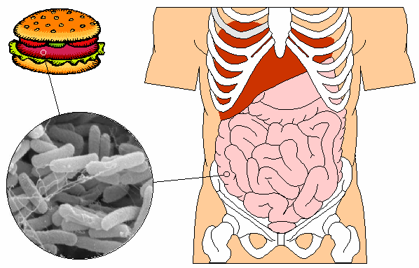
Bacillary dysentery (细菌性痢疾) is the textbook invasive bacterial diarrhoea — the mirror image of cholera (CM exam tested). Where cholera is non-invasive secretory, dysentery is invasive inflammatory — *Shigella* actively invades the colonic mucosa, causing mucoid-bloody stool with tenesmus (the hallmark of an invasive/inflammatory diarrhoea). The exam mindset: bloody-mucoid stool + tenesmus + crampy lower abdominal pain + low-grade fever → think bacillary dysentery (vs amoebic dysentery = subacute, no fever, flask-shaped ulcers). S. dysenteriae serotype 1 is the most severe — produces Shiga toxin and can trigger haemolytic uraemic syndrome (HUS). Diagnosis: stool culture on selective media (XLD, SS agar); treatment: fluoroquinolone (ciprofloxacin) or azithromycin + rehydration.
Core Concepts
1. Definition
Bacillary dysentery = acute invasive bacterial colitis caused by *Shigella* species (or invasive *E. coli*), characterised by fever, abdominal cramps, tenesmus, and mucoid-bloody stool ("痢") from inflammation and ulceration of the sigmoid colon and rectum.
2. Aetiology
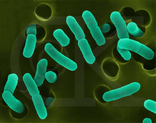
| Feature | Detail |
|---|---|
| Pathogen | *Shigella* — Gram-negative, non-motile rod, facultative intracellular; family *Enterobacteriaceae* |
| Species (4) | S. dysenteriae (serogroup A, most severe, Shiga toxin, epidemic), S. flexneri (B, most common in China), S. boydii (C), S. sonnei (D, mildest, common in industrialised countries) |
| Toxins | Endotoxin (LPS) — fever, toxaemia, DIC, HUS; Shiga toxin (S. dysenteriae 1) — inhibits 60S ribosomal subunit → cell death + endothelial damage → HUS |
| Virulence | Very low inoculum (~10–200 organisms) → highly contagious; survives gastric acid |
| Stability | Sensitive to heat, drying, common disinfectants; survives in cold food/water for days |
3. Epidemiology
- Sporadic and epidemic; seasonal — summer and autumn peak in China and developing countries.
- Common in developing countries with poor water/sanitation; daycare centres, institutions, refugee camps.
- Three links of the chain:
| Link | Detail |
|---|---|
| Source | Patients with acute or chronic dysentery + carriers (especially atypical, chronic, asymptomatic carriers — these are critical for transmission) |
| Route | Faecal-oral — contaminated food, water, vegetables, fruits, daily-use articles; life-contact (hand-to-mouth, fomites) |
| Susceptible | General susceptibility, transient immunity; no cross-immunity between serotypes → repeated infections common |
4. Pathogenesis
- Ingestion → survive gastric acid (very low inoculum needed).
- Adherence and invasion of sigmoid colon and rectal epithelium — *Shigella* is highly invasive (uses Type III secretion system to inject Ipa proteins into host cells → actin rearrangement → macropinocytosis → intracellular survival).
- Intracellular multiplication in epithelial cells + escape into lamina propria.
- Cell-to-cell spread (via IcsA/VirG → actin comet tail) → spreading infection.
- Cell death + endotoxin release → mucosal inflammation, fibrinopurulent exudate, superficial ulceration, necrosis (especially in sigmoid colon and rectum).
- Bleeding + mucopurulent exudate → mucoid-bloody stool + tenesmus (rectal inflammation).
- Endotoxin → systemic inflammation → fever, toxaemia; in severe cases → DIC, HUS, shock.
> HUS (haemolytic uraemic syndrome) = Shiga toxin binds Gb3 on endothelial cells (especially renal) → microangiopathic haemolytic anaemia + thrombocytopenia + AKI. Mostly in children with *S. dysenteriae* 1.
5. Pathological features
- Fibrinous diffuse exudative inflammation in colon (especially sigmoid and rectum).
- Pseudomembranes may form (less classic than *C. difficile*).
- Superficial mucosal necrosis and ulceration → bleeding.
- Most pathology is superficial — full-thickness invasion is rare.
6. Clinical features
Incubation: average 1–3 days (range 12 h to 8 days).
Acute (≤2 months):
| Feature | Detail |
|---|---|
| Fever | Acute onset, 38–40 °C, may be accompanied by chills; in children → febrile seizures |
| Abdominal pain | Crampy, lower abdomen; tenesmus (urge to defecate with little stool — characteristic of rectal involvement) |
| Diarrhoea | Initially watery → mucoid-bloody ("红白痢") within hours–days; 10–30+ stools/day in severe |
| Toxaemia | Headache, malaise, myalgia |
| Dehydration | Variable (less severe than cholera because stool volume is smaller) |
Severe / toxic (中毒型) — more common in children 2–7 years:
- Sudden high fever, convulsions, altered mental status, shock, DIC; paradoxically minimal GI symptoms initially ("无腹泻型") — high mortality.
Chronic (>2 months): persistent loose stools, intermittent mucoid-bloody stools, weight loss, anaemia; often misdiagnosed as IBD.
7. Complications
| Complication | Notes |
|---|---|
| HUS | Microangiopathic haemolytic anaemia + thrombocytopenia + AKI; mostly with *S. dysenteriae* 1 in children; ↑ Cr, ↓ Hb, schistocytes, ↓ platelets |
| Reactive arthritis | Post-infectious, especially HLA-B27+ |
| Seizures | In children, from fever or toxin |
| Toxic megacolon | Rare, severe |
| Bowel perforation | Rare |
| Hypoglycaemia | Children |
| Hypoproteinaemia | Chronic disease |
8. Laboratory examination
| Test | Detail |
|---|---|
| Stool — gross | Mucoid, bloody, small volume; alkaline; pus cells + RBCs |
| Stool microscopy | Many neutrophils (pus cells) + RBCs — distinguishes invasive from secretory diarrhoea |
| Stool culture | XLD agar (red colonies, no H₂S); SS agar; further serotyping with anti-Shigella sera |
| Blood | Leukocytosis; in HUS — schistocytes, ↓ Hb, ↓ platelets, ↑ Cr, ↓ haptoglobin |
| PCR | *ipaH* or *virA* gene — sensitive, rapid |
| Imaging | Abdominal X-ray (toxic megacolon, perforation) |
9. Diagnosis
Clinical + stool microscopy (pus cells + RBCs) + stool culture on XLD/SS. Differential:
| Mimic | Distinguishing |
|---|---|
| Amoebic dysentery (*Entamoeba histolytica*) | Subacute, no fever (usually), flask-shaped ulcers, trophozoites with ingested RBCs in fresh stool |
| EHEC / STEC (*E. coli* O157:H7) | Haemorrhagic colitis + HUS risk; undercooked beef; sorbitol-MacConkey agar |
| Campylobacter, Salmonella, Yersinia | Culture; food exposure |
| Inflammatory bowel disease | Chronic, no fever (usually), negative cultures, endoscopy shows continuous vs skip lesions |
| Intussusception / ischaemic colitis | Older patients, imaging |
| COVID-19 / HIV enteritis | Context |
10. Treatment
| Setting | Regimen |
|---|---|
| Rehydration | ORS (most cases); IV fluids if severe; early refeeding |
| Antibiotics — empirical first-line | Ciprofloxacin 500 mg bid × 3 days (adult); OR azithromycin 500 mg day 1, then 250 mg × 4 days (children, fluoroquinolone resistance, pregnancy); ceftriaxone alternative |
| Avoid | Antidiarrhoeal agents (loperamide, diphenoxylate) — prolong shedding and ↑ risk of HUS |
| HUS | Supportive — fluid balance, dialysis if AKI, blood transfusion if severe anaemia, platelet transfusion only if bleeding or pre-procedure; eculizumab in select cases |
| Chronic | Same antibiotic choice × 7–10 days; check for resistant serotype |
> Resistance watch: fluoroquinolone resistance is rising globally — azithromycin preferred in children and where resistance is high.
11. Prevention
- Hand hygiene — soap and water for 20 s after defecation, before eating; alcohol gel less effective against bacterial spores but adequate for *Shigella*.
- Safe water, sanitation, food hygiene — cook seafood, wash raw vegetables in safe water, refrigerate food.
- Avoid suspected cases preparing food.
- No vaccine currently.
- Antibiotic prophylaxis for close contacts — not routinely recommended (resistance); can consider for institutional outbreaks.
- Notification: Class B notifiable disease (24 h) in China.
High-Yield Points
| Topic | Must-remember |
|---|---|
| Pathogen | *Shigella* — S. dysenteriae (most severe, Shiga toxin), S. flexneri (most common in China), S. boydii, S. sonnei (mildest) |
| Inoculum | Very low (10–200) → highly contagious |
| Hallmark | Mucoid-bloody stool + tenesmus + fever |
| Stool microscopy | Pus cells + RBCs → invasive/inflammatory |
| Pathology | Sigmoid colon + rectum — superficial ulceration, fibrinopurulent exudate |
| Toxins | Endotoxin (LPS) — fever, DIC; Shiga toxin (S. dysenteriae 1) — HUS |
| HUS triad | Microangiopathic haemolytic anaemia + thrombocytopenia + AKI |
| Treatment | Ciprofloxacin × 3 days (adult); azithromycin in children / resistance areas; rehydration |
| Avoid | Loperamide / antidiarrhoeals — prolong shedding, ↑ HUS risk |
| Chronic disease | >2 months — weight loss, intermittent bloody stool |
| Severe/toxic type | Children 2–7; sudden fever + seizures/shock + minimal GI symptoms |
| Differential | Amoebic dysentery (subacute, no fever, flask ulcers, trophozoites with RBCs); EHEC (undercooked beef, HUS); IBD (chronic) |
| Notification | Class B (24 h) |
Topic Summary
Bacillary dysentery is an invasive bacterial colitis caused by *Shigella*, with mucoid-bloody stool + tenesmus + fever — the mirror image of cholera's secretory pattern. S. dysenteriae 1 produces Shiga toxin and can trigger HUS (especially in children). The pathology is in the sigmoid colon and rectum — superficial ulceration. Diagnosis: stool microscopy (pus cells + RBCs) + culture on XLD/SS. Treatment: rehydration + ciprofloxacin (adults) or azithromycin (children/resistance); avoid antidiarrhoeals (↑ HUS risk). Prevention: hand hygiene + safe water/sanitation/food hygiene. Class B notifiable disease.
LMCHK OSCE Practice — Student with Bloody Diarrhoea
Station setup: 21-year-old university student presents with 2-day history of low-grade fever, crampy lower abdominal pain, tenesmus, and ~12 small-volume mucoid-bloody stools per day. He shares a dormitory with 6 others; two have milder diarrhoea.
Candidate tasks (8 min):
- Take a focused exposure + contact history (dining hall, shared toilets, travel, food).
- State the most likely diagnosis (bacillary dysentery — invasive pattern: fever + tenesmus + mucoid-bloody stool).
- List first-line investigations — stool microscopy (pus cells + RBCs), stool culture on XLD/SS, FBC, electrolytes, renal function, HUS screen (Hb, platelets, schistocytes, haptoglobin, LDH).
- Outline immediate management — rehydration (ORS); ciprofloxacin × 3 days (or azithromycin if local resistance high); avoid loperamide; isolate from food handling; notify close contacts.
- Discuss outbreak control — hand hygiene, food safety, notification to dormitory/campus health service (Class B, 24 h), screen close contacts, environmental cleaning.
Key marking cues:
- Recognises invasive pattern (tenesmus + fever + bloody stool) — distinguishes from secretory (cholera).
- Differentiates from amoebic dysentery (subacute, no fever) and EHEC (undercooked beef, HUS).
- Orders stool microscopy (pus cells + RBCs) — basic but exam-essential.
- Avoids loperamide (explain why: prolongs shedding + ↑ HUS risk).
- Recognises dormitory outbreak — public health action + isolation + notification.