Preparatory Mindset
Sepsis is the #1 killer in critical-care medicine worldwide — 50 million cases and 11 million deaths annually (WHO 2018) (CM exam tested). It is the clinical syndrome that ties together every chapter in this ID syllabus: any of viral hepatitis, EHF, dengue, brucellosis, malaria, scrub typhus, cholera, dysentery, or HIV can present with or progress to sepsis. The exam mindset: infection + organ dysfunction = sepsis (Sepsis-3, 2016). qSOFA ≥2 is the rapid bedside screen; SOFA ≥2 confirms organ dysfunction. Treatment is a time-critical bundle (Surviving Sepsis Campaign): lactate → cultures → broad-spectrum antibiotics within 1 h → fluid resuscitation → vasopressors if needed. Every hour of delay in antibiotics increases mortality.
Core Concepts
1. Definition (Sepsis-3, 2016)
> Sepsis = life-threatening organ dysfunction caused by a dysregulated host response to infection. > Septic shock = a subset of sepsis with circulatory and cellular/metabolic abnormalities profound enough to increase mortality; operationalised as vasopressor requirement to maintain MAP ≥65 mmHg + serum lactate >2 mmol/L despite adequate fluid resuscitation.
The shorthand: Sepsis = Infection + Organ Dysfunction.
2. Diagnosis — SOFA and qSOFA
| Score | Use | Components |
|---|---|---|
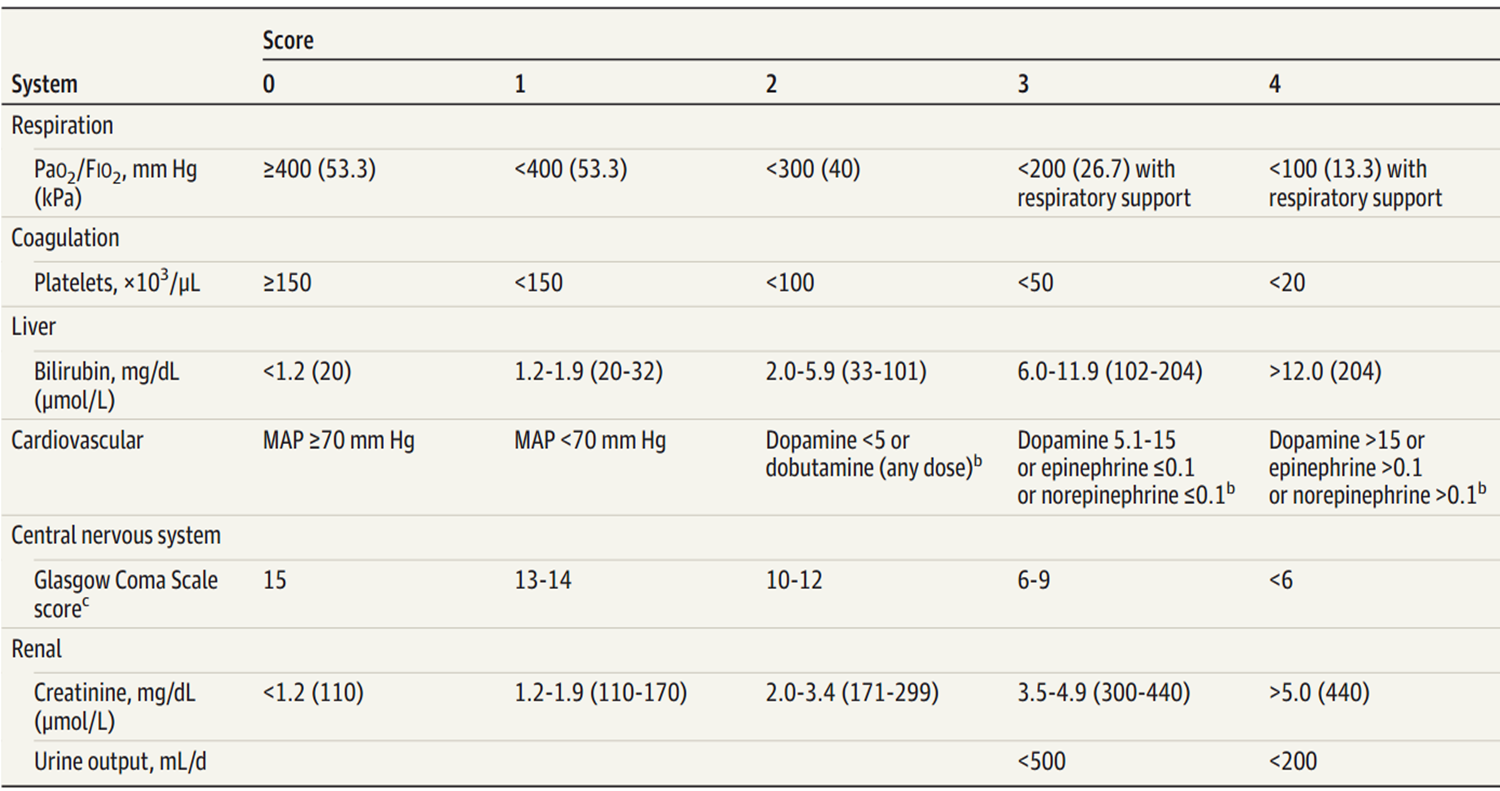
| SOFA (Sequential Organ Failure Assessment) | ICU / ward; ≥2 = organ dysfunction = sepsis | PaO₂/FiO₂, platelets, bilirubin, MAP/vasopressors, GCS, creatinine/urine output |
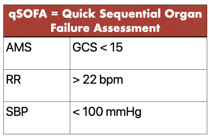
| qSOFA (quick SOFA) | Bedside screen; ≥2 = likely sepsis, escalate care | AMS (GCS <15); RR ≥22; SBP ≤100 mmHg |
3. Epidemiology
- >50 million cases/year worldwide; 11 million deaths (WHO).
- 15 per 1000 hospitalised patients develop sepsis during admission.
- Susceptible populations: ≥65 years, ≤1 year, immune deficiency, severe comorbidities, invasive devices, recent surgery / transplant, pregnancy.
4. Pathogenesis
A cascade:
- Pathogen entry → innate immune activation (TLRs, PAMPs) → cytokine storm (TNF-α, IL-1, IL-6, IL-8) → systemic inflammation.
- Endothelial activation/damage → capillary leak, vasodilation, hypovolaemia, microvascular thrombosis.
- Coagulopathy → DIC (consumption of clotting factors, thrombocytopenia, microthrombi).
- Mitochondrial dysfunction → cellular metabolic failure despite adequate oxygen delivery (cytopathic hypoxia).
- Organ hypoperfusion + direct inflammatory injury → organ dysfunction (lungs — ARDS, kidneys — AKI, brain — encephalopathy, liver — ischaemic hepatitis, heart — septic cardiomyopathy).
5. Clinical manifestations
| System | Finding |
|---|---|
| General | Fever or hypothermia (elderly often hypothermic), rigors, malaise |
| CV | Tachycardia, hypotension, warm peripheries early → cold/mottled late (shock), ↓ capillary refill, narrowed pulse pressure |
| Resp | Tachypnoea, hypoxaemia (ARDS), bilateral crackles if pulmonary oedema |
| Renal | Oliguria, AKI (rising Cr) |
| Hepatic | Jaundice, transaminitis (ischaemic hepatitis), coagulopathy |
| CNS | Confusion, agitation, lethargy, GCS drop |
| Skin | Petechiae, purpura (meningococcemia, DIC), cellulitis at source |
| GI | Ileus, stress ulcer bleeding |
| Lab | WBC ↑ or ↓, neutrophilia + left shift, thrombocytopenia, ↑ lactate (>2), ↑ CRP/PCT, metabolic acidosis, ↑ bilirubin, ↑ Cr, low albumin |
6. Investigations
| Tier | Tests |
|---|---|
| Pathogen | Blood cultures × 2 sets BEFORE antibiotics (during fever spike or chill); urine culture; sputum culture; wound/tissue culture; PCR for culture-negative (16S rRNA broad-range, viral panels); β-D-glucan, galactomannan for fungal sepsis; resin-based media if already on antibiotics |
| Inflammatory | WBC + differential; procalcitonin (PCT) — guides antibiotic duration; CRP — serial trend; IL-6, lactate, ferritin |
| Organ function | LFT, renal function, electrolytes, ABG, lactate, coagulation (PT, aPTT, fibrinogen, D-dimer) |
| Imaging | CXR (pneumonia, effusion, ARDS); abdominal US (abscess); echocardiography (endocarditis); CT chest/abdomen/pelvis (source hunt); CT angiography (vascular complications) |
> Timing of blood cultures: draw immediately before the fever spike or during chills to capture circulating pathogens. If patient is already on antibiotics, resin-based media can absorb residual antibiotics and recover organisms.
7. Differential diagnosis
| Mimic | Clues to distinguish |
|---|---|
| Non-infectious SIRS (pancreatitis, burns, trauma) | No clear infection source; PCT often normal |
| Viral infections (EBV, CMV, dengue, HIV) | Leukopenia + atypical lymphocytes; travel/exposure; PCT normal-to-mildly elevated |
| Haematologic malignancy (leukemia, lymphoma) | Peripheral smear blasts / atypical cells; bone marrow biopsy; PCT normal |
| Adult-Onset Still's Disease | Quotidian spiking fevers, evanescent salmon rash, arthritis; ferritin very high; improves with steroids, not antibiotics |
8. Complications
- Septic shock — vasopressor-requiring hypotension + lactate >2 despite fluid.
- DIC — widespread microvascular thrombosis and bleeding (consume platelets, fibrinogen; prolong PT/aPTT; ↑ D-dimer).
- MODS — progressive dysfunction of ≥2 organs (lungs, kidneys, liver, brain, coagulation).
- ARDS — refractory hypoxaemia from non-cardiogenic pulmonary oedema.
9. Treatment — Surviving Sepsis Campaign (SSC) 1-hour bundle
| Step | Action | Timing |
|---|---|---|
| 1 | Measure lactate; re-measure if >2 | At presentation |
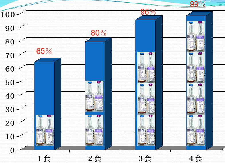
| 2 | Obtain blood cultures BEFORE antibiotics | Before antibiotics |
| 3 | Administer broad-spectrum antibiotics (within 1 h of recognition) | ≤1 h |
| 4 | Begin rapid administration of 30 mL/kg crystalloid for hypotension or lactate ≥4 | Within 3 h |
| 5 | Vasopressors if hypotensive during/after fluid resuscitation to maintain MAP ≥65 mmHg | As needed |
Antibiotic choice — empirical, cover likely source; narrow once culture data returns:
| Suspected source | Empiric regimen |
|---|---|
| Pulmonary (CAP) | β-lactam + macrolide OR respiratory fluoroquinolone |
| Pulmonary (HAP/VAP) | Anti-MRSA + anti-pseudomonal β-lactam |
| Abdominal | Anti-pseudomonal β-lactam + metronidazole (+/− fluconazole) |
| Urinary | Anti-pseudomonal β-lactam (or carbapenem if ESBL risk) |
| Skin/soft tissue | Vancomycin + piperacillin-tazobactam (consider necrotising → surgical consult) |
| Catheter-related | Vancomycin + gram-negative cover; remove/change line |
| Unknown source | Carbapenem ± vancomycin ± antifungal |
Source control — drain abscess, debride necrotic tissue, remove infected device. Delay source control → worse outcome.
Adjuncts:
- Vasopressor: norepinephrine first-line; add vasopressin if refractory.
- Hydrocortisone if refractory shock despite vasopressors.
- IVIG — not routine; consider in toxic shock.
- Blood products: transfuse if Hb <7 g/dL (or <8 in CHD); platelets <20 × 10⁹/L or <50 if active bleeding or procedure.
- Renal replacement therapy for AKI with refractory fluid overload / hyperkalaemia / uraemia.
- Lung-protective ventilation for ARDS (tidal volume 6 mL/kg ideal body weight, plateau pressure ≤30 cmH₂O).
- Glycaemic control target 7.8–10 mmol/L.
- Stress ulcer prophylaxis (PPI) in mechanically ventilated or coagulopathic patients.
- VTE prophylaxis (LMWH unless contraindicated).
10. Prevention
- Infection prevention — hand hygiene, sterile technique, early removal of invasive devices, antibiotic stewardship.
- Vaccination — pneumococcal, influenza, Hib, meningococcal in at-risk populations.
- Screening — early-warning scores (NEWS2, MEWS) on wards to recognise deterioration.
High-Yield Points
| Topic | Must-remember |
|---|---|
| Definition | Sepsis = infection + organ dysfunction (Sepsis-3, 2016); Septic shock = vasopressor-dependent + lactate >2 |
| qSOFA | AMS, RR ≥22, SBP ≤100 mmHg — ≥2 = likely sepsis |
| SOFA | ICU score; ≥2 = organ dysfunction |
| Timing | Antibiotics within 1 h of recognition |
| Cultures | Before antibiotics; 2 sets; during fever spike |
| Fluid | 30 mL/kg crystalloid for hypotension or lactate ≥4 |
| Vasopressor | Norepinephrine first; target MAP ≥65 mmHg |
| Lactate | Measure at presentation; remeasure if >2 |
| Source control | Drain, debride, remove devices — delay = death |
| Differential | AOSD (ferritin ↑↑), hematologic malignancy (PCT normal), viral sepsis (atypical lymphocytes) |
Topic Summary
Sepsis is infection + organ dysfunction — a clinical emergency with mortality rising every hour antibiotics are delayed. Recognise by qSOFA ≥2 at the bedside (AMS, RR ≥22, SBP ≤100), confirm with lactate, blood cultures, and source hunt. Treatment is the SSC 1-hour bundle: measure lactate → cultures → broad-spectrum antibiotics within 1 h → 30 mL/kg crystalloid → norepinephrine to MAP ≥65. Source control (drainage, debridement, device removal) is essential. Complications (shock, DIC, MODS, ARDS) drive mortality; aggressive ICU-level care improves outcome. Prevention is infection control + vaccination + early-warning scores.
LMCHK OSCE Practice — Suspected Sepsis in the Emergency Department
Station setup: 68-year-old diabetic male, day 3 post-op laparotomy, now confused and febrile. T 39.3 °C, HR 128, RR 26, BP 88/52 (MAP 64), SpO₂ 94% on 4 L NP. Urine output 20 mL/h last 2 h. Abdomen soft but wound erythematous with serous discharge.
Candidate tasks (8 min):
- State the most likely diagnosis and explain the criteria met (Sepsis-3).
- List the immediate investigations to order and the order of priority (lactate, blood cultures, FBC, U&E, LFT, ABG, CRP/PCT, wound swab, urinalysis, CXR).
- Outline the first-hour management bundle (30 mL/kg crystalloid, broad-spectrum antibiotics, cultures before antibiotics, MAP target 65).
- Discuss source control (open wound, examine for intra-abdominal collection — CT if stable).
- Explain to the nurse what monitoring to perform (vital signs q15min, urine output hourly, repeat lactate at 2–4 h).
Key marking cues:
- Identifies sepsis from infection (wound) + organ dysfunction (hypotension + oliguria + AMS); qSOFA = 3 (AMS, RR 26, SBP 88).
- Draws blood cultures before antibiotics; chooses empirical cover for abdominal source (e.g., piperacillin-tazobactam ± vancomycin).
- Starts 30 mL/kg crystalloid within 3 h; reassesses MAP.
- Recognises MAP target ≥65; starts norepinephrine if MAP <65 after fluid.
- Notifies surgical team for source control (open wound, possible intra-abdominal collection).
- Avoids prophylactic antibiotics alone without source control.