Preparatory Mindset
Clinical imaging is a case-discussion elective that builds on the systematic Y4S1 Medical Imaging notes — here the exam skill is pattern recognition on real cases. The mindset: for every case, run the same 4-step loop — (1) what does the image show (density/signal/morphology), (2) where is it (organ/system), (3) what is the differential (benign vs malignant, acute vs chronic), (4) what imaging sign clinches it. New advances (dual-source CT, dual-energy, photon-counting, AI, tomosynthesis, MRI/PET fusion) are a high-yield exam topic because they ask "which modality/technology for which clinical question". The LMCHK angle: radiology OSCE stations ask you to read a film, name the sign, and give the next step — exactly the case-discussion format of this elective.
Core Concepts
1. The new advances — why they matter
| Technology | What it does | Clinical value |
|---|---|---|
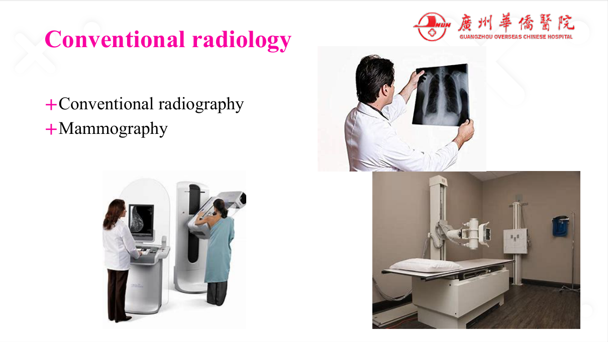
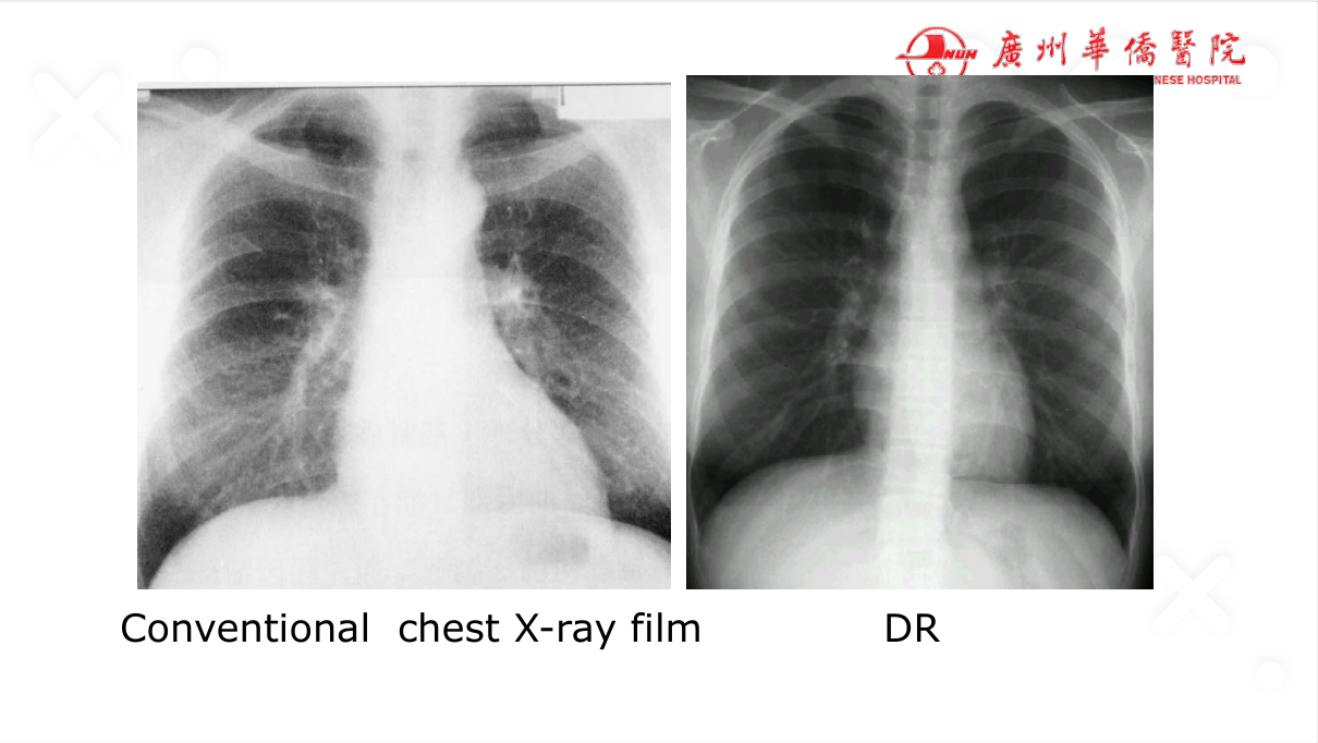
| Digital radiography (DR) | Replaces film; digital processing | Higher contrast sensitivity, lower dose, faster, machine-aided (CAD) diagnosis |
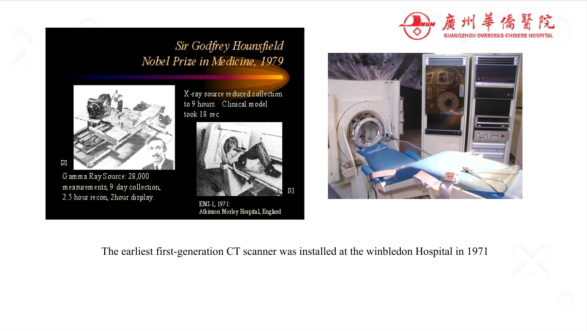
| Dual-source / dual-energy CT | Two imaging chains mounted at angles; separate energy spectra | Spectral material decomposition (e.g., virtual non-contrast, bone removal, uric acid detection), faster scanning, lower contrast dose |
| Photon-counting CT | Detector counts individual photons with energy discrimination | Higher spatial/contrast resolution, lower radiation, material-specific imaging |
| Spectral / multi-energy CT | Material decomposition (iodine, calcium, uric acid) | Better lesion characterisation (e.g., gout vs pseudo-gout, thrombus vs calcification) |
| AI / deep learning (CAD) | Automated lesion detection, triage, quantification | Timely diagnosis for patients who may otherwise wait — nodule detection, intracranial haemorrhage triage, mammography |
| Digital breast tomosynthesis (DBT) | 3D mammography — slice reconstruction | Reduces tissue overlap — improves cancer detection, reduces recall |
| Quantitative imaging | Automated measurement (volumes, densities, perfusion) | Standardised follow-up (e.g., RECIST, tumour response) |
| Advanced MRI (diffusion, perfusion, spectroscopy, DTI) | Functional + structural | Tumour grading (ADC), ischaemia (DWI), tractography (DTI) |
| PET/MRI, PET/CT fusion | Metabolic + anatomical | Tumour staging, therapy response |
2. The case format — how to read a case discussion
Each case in this elective follows the same structure, which is exactly what you will be tested on:
- Clinical presentation — age, sex, symptoms (e.g., chest pain for half a month).
- Imaging findings — describe the lesion systematically (location, size, borders, density/signal, enhancement, effect on surroundings).
- Differential diagnosis — rank by likelihood using imaging + clinical features.
- Diagnosis + next step — the definitive imaging/management path.
> Exam pearl: always describe the lesion with location → morphology → density/signal → enhancement → surrounding changes — the "read the film" OSCE structure.
3. When to choose which modality (the classic pairing questions)
| Clinical question | First-line / best modality |
|---|---|
| Acute chest pain (rule out dissection/PE) | CT (CTPA / CT aortogram) — fast, high accuracy |
| Focal liver lesion characterisation | Contrast-enhanced ultrasound (CEUS) or MRI with contrast (hemangioma = MRI pathognomonic) |
| Breast cancer screening | Mammography (+ tomosynthesis) |
| Bone tumour characterisation | MRI (marrow involvement) + plain film (matrix/mineralisation) |
| Acute stroke (rule out haemorrhage → thrombolysis) | NCCT (bleed) then CTA/MRI-DWI (ischaemia) |
| Appendicitis | Ultrasound (children/pregnancy) or CT (adults) |
| Focal liver lesion — haemangioma vs HCC | MRI (haemangioma: T2 bright, peripheral nodular enhancement; HCC: arterial enhancement + washout) |
High-Yield Points
| Topic | Must-remember |
|---|---|
| Dual-energy CT | Material decomposition (iodine/bone/urate) + virtual non-contrast |
| Photon-counting CT | Energy-discriminating detector — higher resolution, lower dose |
| AI in radiology | Automated detection/triage — timely diagnosis |
| Tomosynthesis | 3D mammography — reduces overlap, better detection |
| Film-reading structure | Location → morphology → density/signal → enhancement → surroundings |
| Case format | Presentation → findings → differential → diagnosis → next step |
| MRI lesion characterisation | DWI (ischaemia/tumour grade), ADC, enhancement pattern |
| PET/CT fusion | Metabolic + anatomical staging |
| CEUS | Real-time contrast imaging of focal liver lesions |
Topic Summary
This elective is case-discussion based — every lecture presents real cases to teach film-reading and differential diagnosis. The new-advances lecture covers dual-source/dual-energy CT (material decomposition), photon-counting CT, AI-assisted detection (timely diagnosis), tomosynthesis (3D mammography), quantitative imaging, and MRI/PET fusion. The transferable skill is the systematic film-reading loop (location → morphology → density/signal → enhancement → surroundings) and modality selection for the clinical question — the exact competencies tested in LMCHK film-reading OSCE stations.
LMCHK OSCE Practice — Choosing the Right Imaging Test
Station setup: A 58-year-old man presents with acute severe tearing chest pain radiating to the back, BP 180/100 in the right arm, 120/70 in the left. You suspect aortic dissection.
Candidate tasks (8 min):
- Recognise the clinical picture of acute aortic syndrome (tearing pain + BP differential).
- State the best imaging test — CT aortogram (CT angiography) — fast, definitive; (alternatives: TOE/MRI if CT unavailable/contraindicated).
- Explain what CT shows: intimal flap, true/false lumen, extent of dissection, involvement of arch vessels/pericardium.
- Mention the role of dual-source/dual-energy CT (fast, low dose, virtual non-contrast — can subtract calcium).
- Outline immediate management: BP control (β-blocker), analgesia, cardiothoracic surgical referral for type A; imaging must not delay transfer.
Key marking cues:
- CTA is the test of choice for dissection.
- Knows the Stanford classification (type A = ascending — surgery; type B = descending — medical).
- Uses dual-energy CT's virtual non-contrast concept.
- Balances imaging speed vs clinical urgency.