Preparatory Mindset
Liver imaging (174-page case discussion) is the focal liver lesion chapter the single highest-yield abdominal imaging topic. The mindset: characterise the lesion by enhancement pattern across phases (arterial, portal venous, delayed) using CEUS/CT/MRI: the big four hemangioma (peripheral nodular centripetal fill, persistent), HCC (arterial hyperenhancement + washout in a cirrhotic liver), FNH (homogeneous arterial enhancement + central scar, normal liver), metastasis (hypovascular or rim enhancement + washout). The exam pearl: in a cirrhotic liver, any enhancing lesion with washout = HCC until proven otherwise (surveillance: US + AFP q6 months). The LMCHK angle: "incidental liver lesion" benign vs malignant characterisation + the cirrhotic screening pathway.
Core Concepts
1. The four-phase approach to focal liver lesions
Phases (CT/MRI with contrast): unenhanced arterial (25-35 s) portal venous (60-70 s) delayed (3-5 min).
| Lesion | Arterial | Portal venous / delayed | Key features |
|---|---|---|---|
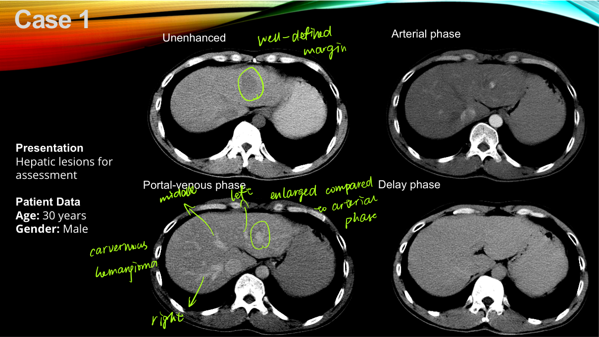
| Hemangioma (HH) most common benign | Peripheral nodular enhancement | Progressive centripetal fill persistent (isodense to blood on delayed) | Cavernous; well-defined; MRI T2 very bright ("light bulb"); no cirrhosis association |
| HCC | Arterial hyperenhancement (intense) | Washout (hypo on portal/delayed) + capsule (delayed rim) | Cirrhotic liver, AFP ; mosaic/necrosis; portal vein invasion (tumour thrombus) |
| FNH | Homogeneous hyperenhancement + spoke-wheel/stellate artery | Iso/hyper (stays bright), central scar enhances on delayed | Normal liver, young woman, no capsule, no washout |
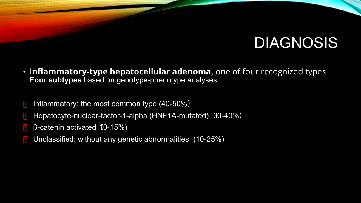
| Hepatocellular adenoma (HCA) | Arterial enhancement | Iso/washout variable | OCP (young woman), haemorrhage risk, malignant transformation ( -catenin); fatty |
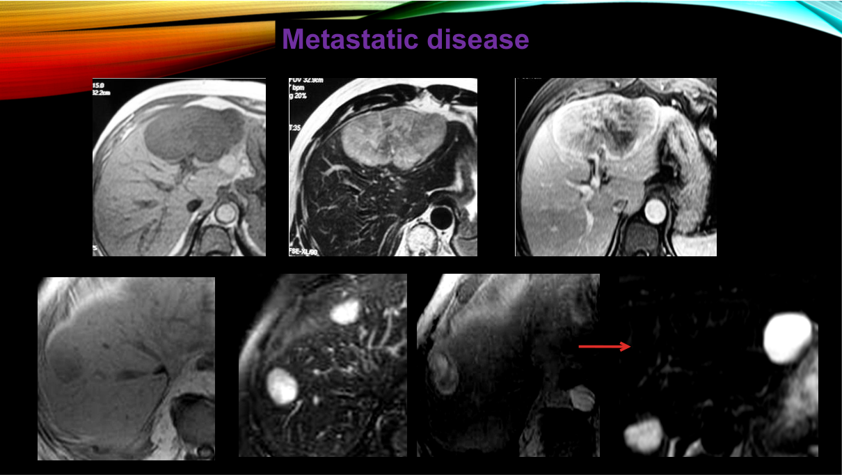
| Metastasis | Rim/peripheral enhancement (hypovascular common: colon) or hyper (NET/renal/thyroid) | Washout | Multiple lesions, known primary, "target sign" (ring + central) |
| Cyst | No enhancement | No enhancement | Simple anechoic/water density, thin wall |
| Abscess | Rim enhancement | Persistent rim, surrounding oedema | Fever, painful, gas possible (pyogenic), cluster sign |
| Cholangiocarcinoma | Peripheral rim | Delayed progressive enhancement (fibrosis) | Intrahepatic mass with capsular retraction; CA19-9 |
2. HCC the cirrhotic screening and diagnosis
Risk: cirrhosis (HBV/HCV China), alcohol, NAFLD. Surveillance: abdominal US + AFP every 6 months in at-risk patients.
Diagnosis (non-invasive, LI-RADS): in a cirrhotic liver, a nodule 1 cm with arterial hyperenhancement + washout (+ enhancing capsule) on MRI/CT (LI-RADS 5) = HCC biopsy not required. Portal vein tumour thrombus = advanced.
Staging/treatment: BCLC staging very early/early (single small) resection / ablation (RFA) / transplant (Milan criteria); intermediate (multinodular) TACE (chemoembolisation the IR chapter's mainstay); advanced (portal invasion/metastatic) systemic (sorafenib/lenvatinib + immunotherapy).
3. Hemangioma vs FNH vs HCC the table that wins exams
| Feature | Hemangioma | FNH | HCC |
|---|---|---|---|
| Liver background | Normal | Normal | Cirrhotic |
| Patient | Any | Young woman | Cirrhotic (M>F) |
| Arterial | Peripheral nodular | Homogeneous + spoke-wheel | Hyperenhancement |
| Delayed | Persistent (fill) | Central scar enhances | Washout + capsule |
| Biopsy needed? | No (typical) | No (typical) | No (LI-RADS 5) |
| Danger | None | None | Malignant |
4. Other liver cases
- Liver metastasis workup: CT chest/abdomen/pelvis + PET-CT; biopsy if no primary found.
- Liver abscess: pyogenic (portal seeding, biliary) vs amoebic (right lobe, "anchovy paste"); drainage + antibiotics; US/CT guided.
- Portal hypertension / cirrhosis imaging: nodular liver, splenomegaly, ascites, varices, cavernous transformation of portal vein; Budd-Chiari (hepatic vein thrombosis).
- Focal fatty change / focal sparing: geographic, no mass effect, no vascular displacement.
- Cystic lesions: simple cyst vs hydatid (echinococcus China: calcified cyst, daughter cysts) vs cystadenoma/cystadenocarcinoma.
High-Yield Points
| Topic | Must-remember |
|---|---|
| Big four liver lesions | Hemangioma, HCC, FNH, metastasis |
| Hemangioma | Peripheral nodular centripetal fill, persistent; T2 "light bulb" |
| HCC | Arterial hyperenhancement + washout + capsule (cirrhotic liver) |
| HCC surveillance | US + AFP q6 months (cirrhosis) |
| HCC non-invasive Dx | LI-RADS 5 biopsy not required in cirrhosis |
| FNH | Central scar (enhances delayed) + spoke-wheel, young woman, normal liver |
| Adenoma | OCP, haemorrhage, malignant potential |
| Metastasis | Multiple, rim enhancement + washout, target sign |
| Abscess | Rim + fever; drainage + antibiotics |
| Cholangiocarcinoma | Delayed progressive enhancement + capsular retraction; CA19-9 |
| Hydatid | Calcified cyst + daughter cysts (echinococcus) |
| HCC treatment | Resection/ablation/transplant (early); TACE (intermediate); systemic (advanced) |
Topic Summary
Liver imaging is focal-lesion characterisation by enhancement pattern: hemangioma (peripheral nodular centripetal fill, persistent), HCC (arterial hyperenhancement + washout + capsule in cirrhosis, LI-RADS 5, no biopsy), FNH (homogeneous + central scar + spoke-wheel, young woman), metastasis (multiple, rim + washout). Cirrhotic livers are screened with US + AFP q6 months; HCC is treated by resection/ablation/transplant (early), TACE (intermediate), systemic therapy (advanced). Abscess (drain), cholangiocarcinoma (delayed enhancement), and hydatid cysts (calcified, China) round out the case set.
LMCHK OSCE Practice Incidental Liver Lesion
Station setup: A 55-year-old man with chronic HBV cirrhosis is on surveillance. US shows a 2.5-cm nodule in the right lobe. Contrast MRI: arterial hyperenhancement with washout on portal venous phase and an enhancing capsule on delayed phase. AFP 200.
Candidate tasks (8 min):
- Interpret: arterial hyperenhancement + washout + capsule in a cirrhotic liver = HCC (LI-RADS 5) no biopsy needed.
- Explain surveillance caught it early (US + AFP q6 months in cirrhosis).
- Stage the disease (single 2.5-cm lesion, no vascular invasion BCLC early) and discuss treatment options: resection, radiofrequency ablation (RFA), or liver transplant (Milan criteria) curative intent.
- Mention TACE if not resectable/multinodular; systemic therapy if advanced (portal invasion).
- Counsel on follow-up (imaging + AFP) and aetiology control (HBV antiviral).
Key marking cues:
- LI-RADS 5 non-invasive diagnosis the key modern concept.
- Correct staging + treatment choice (early HCC curative).
- Knows surveillance protocol (US + AFP q6 months).
- Covers aetiology control (HBV) and follow-up.