Preparatory Mindset
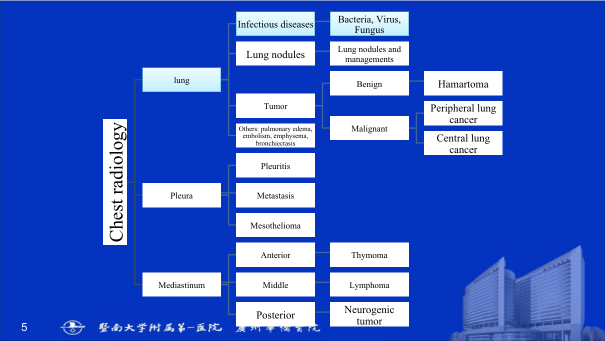
Chest radiology is the highest-yield film-reading topic — it appears in every OSCE and final. The mindset: a systematic 7-point checklist (technique, airways, lungs, pleura, mediastinum, diaphragm, bones/soft tissues) — then the disease patterns: consolidation (air bronchogram), collapse/atelectasis (volume loss, silhouette sign), nodules/masses, pneumothorax, pleural effusion, and the mediastinum (widening, masses). The LMCHK angle: "read this CXR" stations — name the sign, give the diagnosis, state the next step. Chest CT (mediastinum, hila, nodules, HRCT) extends the same patterns. The elective's four chest parts (I-IV) cover: technique + pattern approach, consolidation/collapse, nodules/masses, and the mediastinum.
Core Concepts
1. The systematic approach (7 points)
- Technique — PA? rotation? inspiration (5-7 ribs anterior)? penetration (spines visible)?
- Airway — trachea central? (deviation → collapse/tension/mass)
- Lungs — opacities, lucencies, nodules; compare sides.
- Pleura — effusion, pneumothorax, thickening.
- Mediastinum — width, contour (see part IV).
- Diaphragm — height (right higher than left by ~1-2 cm), flattening (COPD), free air under (perforation).
- Bones/soft tissues — ribs (fracture, notching), spine, shoulders, soft-tissue gas.
2. The patterns (the core of the lecture)
| Pattern | Definition | Causes | Key sign |
|---|---|---|---|
| Consolidation | Airspace filling (pus/fluid/blood/cells) | Pneumonia (lobar), pulmonary oedema, haemorrhage, aspiration | Air bronchogram (air in bronchi against opaque parenchyma), lobar distribution, no volume loss |
| Collapse (atelectasis) | Volume loss | Endobronchial obstruction (tumour, FB, plug), compression, scarring | Volume loss — fissure/mediastinum shift toward the lesion, elevated diaphragm, compensatory hyperinflation; silhouette sign |
| Nodule | <3 cm rounded opacity | Granuloma (TB — most common in China), tumour (primary/metastasis), inflammatory | Size, margins (spiculated = malignant), calcification pattern (benign: central/laminated/popcorn) |
| Mass | >3 cm | Tumour (lung cancer), abscess, aneurysm | Location (central vs peripheral), cavitation, invasion |
| Pneumothorax | Air in pleural space | Spontaneous (tall thin young male), trauma, iatrogenic | Visceral pleural line + absent lung markings peripherally; tension = mediastinal shift + flattened diaphragm (emergency) |
| Pleural effusion | Fluid in pleural space | Transudate (HF, cirrhosis) vs exudate (infection, malignancy, PE) | Blunting of costophrenic angle (300 mL+), meniscus sign; massive = white-out + mediastinal shift away |
| Miliary pattern | Multiple tiny nodules | TB (miliary), metastases, sarcoidosis | Diffuse 1-3 mm nodules |
| Cavity | Air-filled space in a lesion | TB, abscess, lung cancer (cavitating SCC), Wegener | Thick irregular wall (malignancy) vs thin (benign) |
| Hyperlucency | Increased lucency | COPD/emphysema, pneumothorax, bullae | Fewer vessels, flat diaphragms, barrel chest |
3. The silhouette sign
Loss of the normal interface between an opacity and an adjacent structure of similar density tells you where the lesion is:
| Silhouette sign | Location of opacity |
|---|---|
| Right heart border obscured | Right middle lobe |
| Left heart border obscured | Lingula |
| Aortic knuckle obscured | Left upper lobe (apicoposterior) |
| Diaphragm obscured | Lower lobes |
| Cardiac apex lost | Pericardial/effusion vs lower lobe |
4. Lobar collapse patterns (high-yield)
| Lobe | Pattern |
|---|---|
| RUL | Opacification of upper zone, minor fissure elevated ("apex pulled up") |
| LUL | Veil-like opacity, aortic knuckle silhouette sign, "Luftsichel" sign (air crescent) |
| RML | Triangular opacity at right heart border (silhouette sign) |
| RLL | Right lower zone opacity, heart border preserved, fissure down |
| Lingula | Left heart border lost, triangular |
5. The mediastinum (chest part IV)
Mediastinal compartments and masses:
| Compartment | Contents | Masses |
|---|---|---|
| Anterior | Thymus, lymph nodes, fat | Thymoma (most common), lymphoma, teratoma/germ cell, retrosternal thyroid, lymphadenopathy — "the 4 Ts: Thymoma, Terrible lymphoma, Teratoma, Thyroid" |
| Middle | Heart, great vessels, trachea, nodes | Lymphadenopathy (TB/sarcoid/lymphoma), aortic aneurysm, bronchogenic cyst |
| Posterior | Oesophagus, descending aorta, nerves | Neurogenic tumour (schwannoma, neurofibroma — most common), oesophageal lesion, paraspinal abscess/mass |
Mediastinal widening: aortic aneurysm/dissection, lymphoma, thymoma, thyroid; superior mediastinal widening in elderly = aortic unfolding (tortuosity — normal ageing), NOT a mass.
Pneumomediastinum — air outlining mediastinal structures (Boerhaave/oesophageal rupture, trauma, asthma).
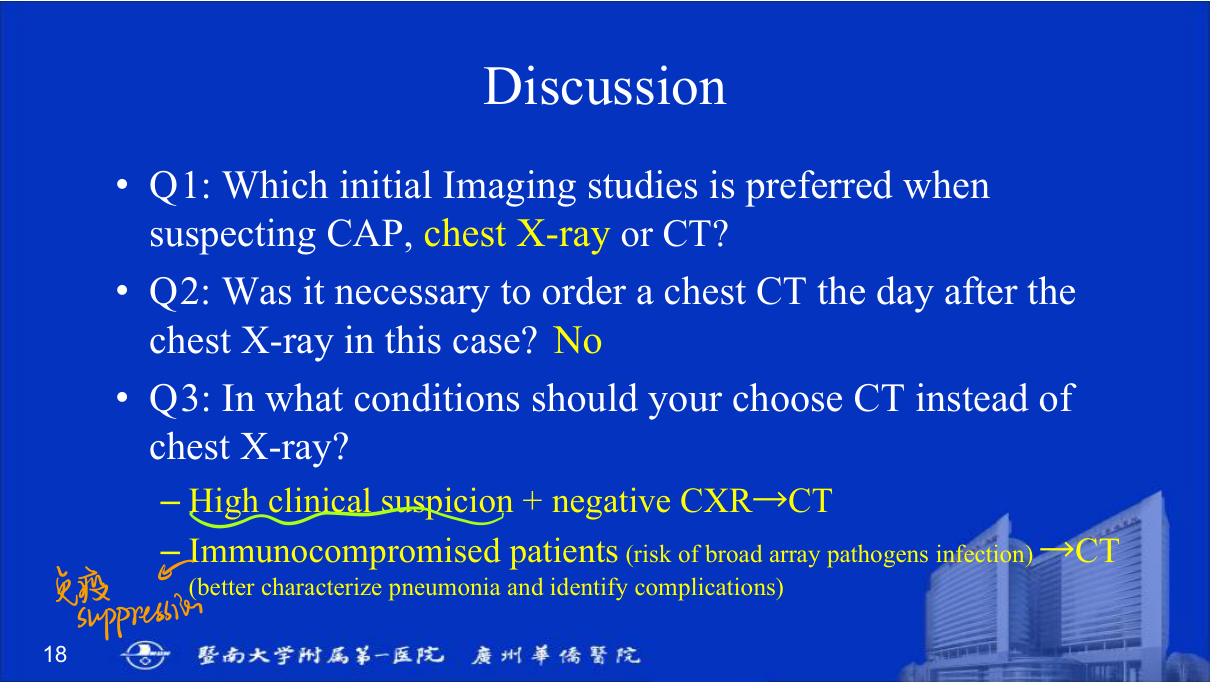
6. Chest CT essentials
- HRCT — interstitial lung disease (reticulation, honeycombing = UIP; ground glass).
- Nodule characterisation — solid vs part-solid/ground-glass (adenocarcinoma spectrum), growth on follow-up.
- Mediastinal/hilar staging — lymph nodes, lung cancer staging (TNM).
- CTPA — PE (see CVS chapter).
High-Yield Points
| Topic | Must-remember |
|---|---|
| 7-point CXR checklist | Technique, airway, lungs, pleura, mediastinum, diaphragm, bones |
| Consolidation | Air bronchogram, no volume loss |
| Collapse | Volume loss, silhouette sign, shift toward lesion |
| Silhouette sign | Right heart = RML; Left heart = lingula; Aorta = LUL |
| Nodule | <3 cm; spiculated = malignant; benign calcification patterns |
| Pneumothorax | Pleural line + no peripheral markings; tension = shift (emergency) |
| Effusion | Blunted CP angle (300 mL+), meniscus; massive = shift away |
| Miliary | TB, mets, sarcoid — 1-3 mm nodules |
| Anterior mediastinum | Thymoma, lymphoma, teratoma, thyroid (4 Ts) |
| Posterior mediastinum | Neurogenic tumour most common |
| HRCT | Reticulation/honeycombing = UIP |
| Part-solid nodule | Adenocarcinoma spectrum — follow up |
Topic Summary
Chest radiology is read systematically (7 points) and by pattern: consolidation (air bronchogram, no volume loss), collapse (volume loss + silhouette sign + shift toward lesion), nodules (<3 cm, spiculated = malignant), masses (>3 cm), pneumothorax (pleural line; tension = emergency), effusion (meniscus; massive = shift away), miliary (TB/mets), cavity, and hyperlucency. The silhouette sign localises (right heart = RML, left heart = lingula, aorta = LUL). Mediastinal compartments predict masses — anterior 4 Ts (thymoma, lymphoma, teratoma, thyroid), posterior neurogenic. Chest CT/HRCT extends the same patterns (nodule characterisation, ILD, staging).
LMCHK OSCE Practice — Read the Chest X-Ray
Station setup: A 60-year-old male smoker presents with haemoptysis. CXR shows a 5-cm right hilar mass with right upper lobe collapse (elevated minor fissure, opacity silhouetting the aortic side? — right upper zone), widened right hilum.
Candidate tasks (8 min):
- Read systematically: technique → airway → lungs → pleura → mediastinum → diaphragm → bones.
- Describe: right hilar mass + RUL collapse (fissure elevated, volume loss) — the classic "S sign of Golden" (reversed S — hilar mass + collapsed upper lobe).
- Differential: lung cancer (SCC/central — most likely in a smoker with haemoptysis), TB, lymphoma.
- Recommend next step: CT chest (staging — lesion, nodes, mets) + bronchoscopy + biopsy for tissue diagnosis; PET-CT for staging.
- Discuss urgency — haemoptysis + central obstructing mass → bronchoscopy for diagnosis and airway assessment.
Key marking cues:
- Systematic reading and names the S sign of Golden (hilar mass + lobar collapse).
- Links smoking + haemoptysis + central mass → lung cancer.
- CT + bronchoscopy + biopsy as the next steps.
- Mentions staging (PET-CT, TNM) and smoking cessation.