Preparatory Mindset
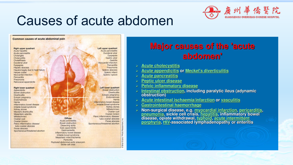
Acute abdomen imaging is the surgical emergency imaging chapter — the mindset is "is this surgical?" The three great questions: (1) is there free air (perforation)? (2) is there obstruction (and where)? (3) is there a surgical inflammatory process (appendicitis, cholecystitis, pancreatitis)? Plain films (erect CXR/decubitus for free air; AXR for obstruction/ileus) → CT is the workhorse for adults (appendicitis, diverticulitis, obstruction, ischaemia); US first in children and pregnancy (appendicitis, gynae). The exam pearls: free air under the diaphragm = perforation until proven otherwise; small bowel obstruction (valvulae conniventes) vs large bowel obstruction (haustra); appendicitis = CT dilated appendix + fat stranding; intussusception = target sign (child, jelly stool).
Core Concepts
1. Free air (pneumoperitoneum) — the perforation question
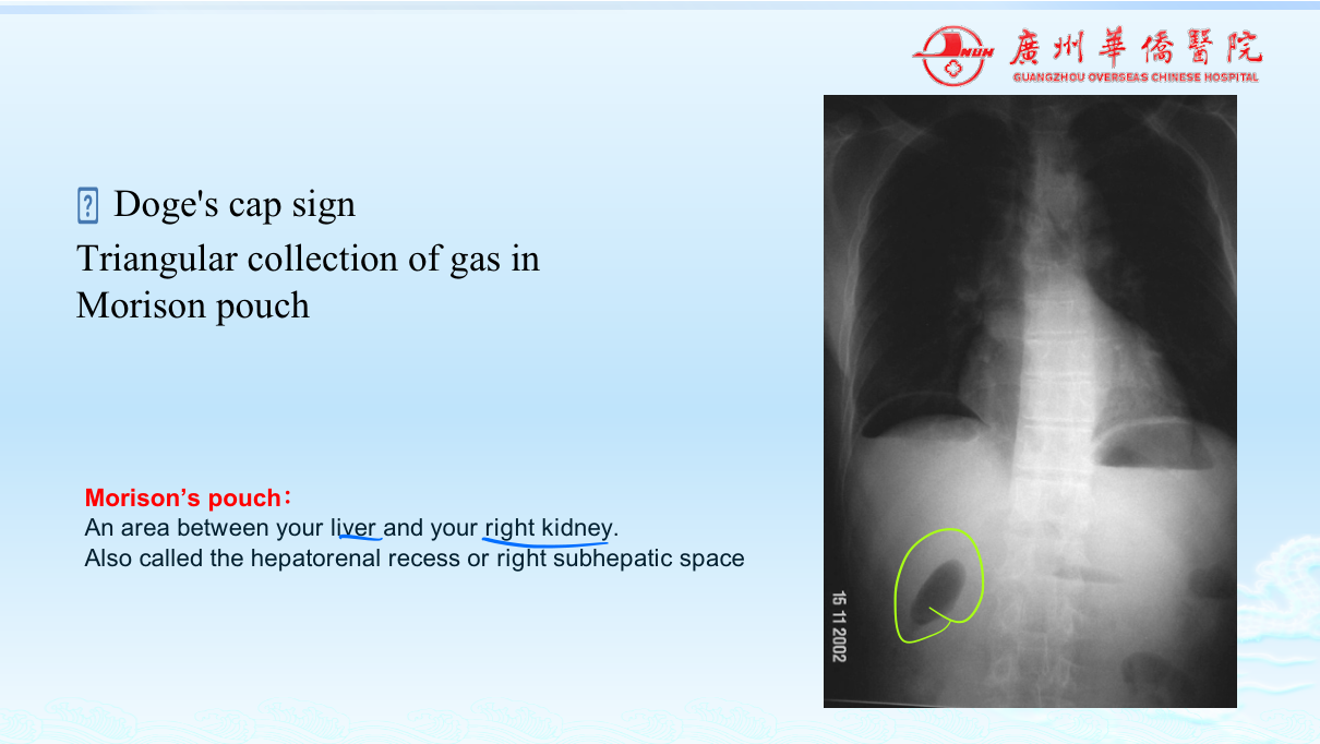
Cause: GI perforation (peptic ulcer, diverticulitis, tumour, trauma, NEC in neonates), post-surgery (24 h+).
Imaging:
- Erect chest X-ray / left lateral decubitus — free air under the diaphragm (crescent lucency).
- CT — the most sensitive: free air (pneumoperitoneum) localised — subphrenic, around liver, small volume.
Clinical correlation: sudden severe abdominal pain, board-like rigidity (perforated ulcer), sepsis. Free air + surgical abdomen → laparotomy (or laparoscopy).
Caveat: CT can detect very small volumes that CXR misses; post-operative air resolves over days — context matters.
2. Bowel obstruction — the "where and why"
| Type | Location | Plain film / CT | Causes |
|---|---|---|---|
| Small bowel obstruction (SBO) | Small bowel | Dilated small bowel loops (central, valvulae conniventes — complete transverse lines), air-fluid levels ("staircase"), collapsed colon | Adhesions (most common — post-surgery), hernia, tumour, Crohn |
| Large bowel obstruction (LBO) | Colon | Dilated peripheral colon (haustra — incomplete lines), ileocaecal valve competence determines small bowel involvement | Tumour (most common), volvulus (sigmoid — "coffee bean sign"), diverticular stricture, faecal impaction |
| Ileus | Non-mechanical | Dilated both small + large bowel, no transition point | Post-op, peritonitis, pancreatitis, metabolic |
| Volvulus | Sigmoid (adult), midgut (child) | "Coffee bean" / "bird beak" (contrast) | Twisting of mesentery — vascular compromise = emergency |
The "transition point" — the site of calibre change — localises the obstruction; CT identifies the cause + closed-loop/ischaemia signs (strangulation — emergency).
3. Surgical inflammatory processes
| Disease | Best imaging | Findings |
|---|---|---|
| Appendicitis | US (child/pregnancy) or CT (adult) | Dilated appendix >6 mm, non-compressible, wall thickening, fat stranding, appendicolith; complications — abscess, perforation (free air) |
| Cholecystitis | US | Gallstones + thickened GB wall (>3 mm), pericholecystic fluid, Murphy sign (US probe), sonographic Murphy; complications — perforation, empyema |
| Pancreatitis | CT (contrast) | Enlarged pancreas, peripancreatic fat stranding, necrosis (non-enhancing), fluid collections, gas (infected necrosis); Balthazar score |
| Diverticulitis | CT | Diverticula + wall thickening + pericolic fat stranding/abscess; Hinchey grading |
| Intussusception (child) | US | "Target/doughnut sign" (transverse), "pseudokidney" (longitudinal) — telescoping bowel; air/pneumatic reduction |
| Mesenteric ischaemia | CT angiography | Vessel occlusion (SMA), bowel wall thickening, pneumatosis (gas in wall), portal venous gas — emergency |
| Bowel ischaemia (strangulation) | CT | Closed-loop, poor enhancement, pneumatosis |
4. The acute abdomen imaging strategy (exam summary)
| Clinical question | First test |
|---|---|
| Perforation? | Erect CXR (free air) → CT if negative |
| Obstruction? | AXR (dilated loops/levels) → CT (transition point + cause) |
| Appendicitis (adult)? | CT |
| Appendicitis (child/pregnancy)? | US |
| Cholecystitis? | US |
| Pancreatitis severity? | Contrast CT |
| Intussusception (child)? | US (target sign) → air enema reduction |
| Gynae (ectopic, torsion, cyst)? | US (transvaginal) |
High-Yield Points
| Topic | Must-remember |
|---|---|
| Free air | Under diaphragm on erect CXR; CT most sensitive — perforation |
| SBO | Valvulae conniventes (transverse lines), central, air-fluid levels; adhesions most common |
| LBO | Haustra (incomplete lines), peripheral; tumour most common |
| Sigmoid volvulus | "Coffee bean sign" |
| Transition point | Localises obstruction; CT finds cause + ischaemia |
| Appendicitis | US (child/preg) or CT (adult) — appendix >6 mm + stranding |
| Cholecystitis | US — stones + GB wall >3 mm + Murphy |
| Pancreatitis | Contrast CT — necrosis (non-enhancement), Balthazar |
| Diverticulitis | CT — diverticula + wall thickening + stranding |
| Intussusception | US target sign → air enema reduction |
| Mesenteric ischaemia | CTA — SMA occlusion, pneumatosis, portal venous gas — emergency |
| Ileus | Both small + large dilated, no transition point |
Topic Summary
Acute abdomen imaging answers three questions: perforation (free air — erect CXR → CT), obstruction (AXR → CT with transition point; SBO valvulae vs LBO haustra; volvulus coffee bean), and surgical inflammation (appendicitis — CT adult/US child; cholecystitis — US; pancreatitis — contrast CT; diverticulitis — CT). Intussusception (US target sign → air reduction) and mesenteric ischaemia (CTA, pneumatosis — emergency) complete the surgical set. The strategy: CXR first for free air, CT for the adult surgical abdomen, US for children/pregnancy/gallbladder/gynae.
LMCHK OSCE Practice — The Acute Abdomen
Station setup: A 24-year-old man presents with central abdominal pain migrating to the right iliac fossa over 12 hours, anorexia, low-grade fever. RIF tenderness with rebound. WBC 14. You are asked about imaging.
Candidate tasks (8 min):
- Recognise the classic appendicitis history (migrating pain, anorexia, RIF tenderness).
- Recommend imaging: US (first-line, especially young — graded compression, appendix >6 mm non-compressible) or CT (more sensitive — dilated appendix + fat stranding) if US equivocal.
- Describe the CT findings (appendix >6 mm, wall thickening, periappendiceal fat stranding, ± appendicolith, ± abscess).
- Discuss complications (perforation — free air/abscess, peritonitis) and why imaging matters (avoid negative appendicectomy).
- Outline management: IV antibiotics + appendicectomy (laparoscopic) — emergency; drain abscess if present.
Key marking cues:
- US first in young patients (radiation), CT if equivocal/adult.
- Knows appendicitis imaging criteria (dilated non-compressible appendix).
- Identifies complications (abscess, perforation).
- Links imaging to surgical decision (appendicectomy).