Preparatory Mindset
Head & neck imaging in this elective is case-based — the classic cases are nasopharyngeal carcinoma (NPC) (a China/Southern-China endemic cancer — the headliner), plus neck masses (thyroid, salivary, lymph nodes) and the imaging questions around them. The mindset: NPC = the "endemic sinus tumour" — unilateral serous otitis media, epistaxis, nasal obstruction, cervical lymphadenopathy, and cranial nerve palsies (VI, V, III) → MRI (the best for soft tissue/tumour extent) + CT (bone erosion) + nasopharyngoscopy + biopsy. The LMCHK angle: "painless neck mass" and "unilateral glue ear in an adult" → think NPC — the imaging workup is MRI + biopsy.
Core Concepts
1. Nasopharyngeal carcinoma (NPC) — the headliner case
Epidemiology: endemic in Southern China (Guangdong, Hong Kong), SE Asia — EBV-associated; peaks 40-60; male predominance.
Clinical (the presentation cluster):
- Unilateral serous otitis media / glue ear in an adult (Eustachian tube obstruction) — "adult with unilateral glue ear = NPC until proven otherwise"
- Epistaxis, blood-stained sputum, nasal obstruction
- Cervical lymphadenopathy (often posterior cervical chain — the "node of Rouvière"/retropharyngeal)
- Cranial nerve palsies — VI (abducens — most common), V (trigeminal), III, IV — via the cavernous sinus
- Headache, trismus (pterygoid invasion)
Imaging:
| Modality | Role |
|---|---|
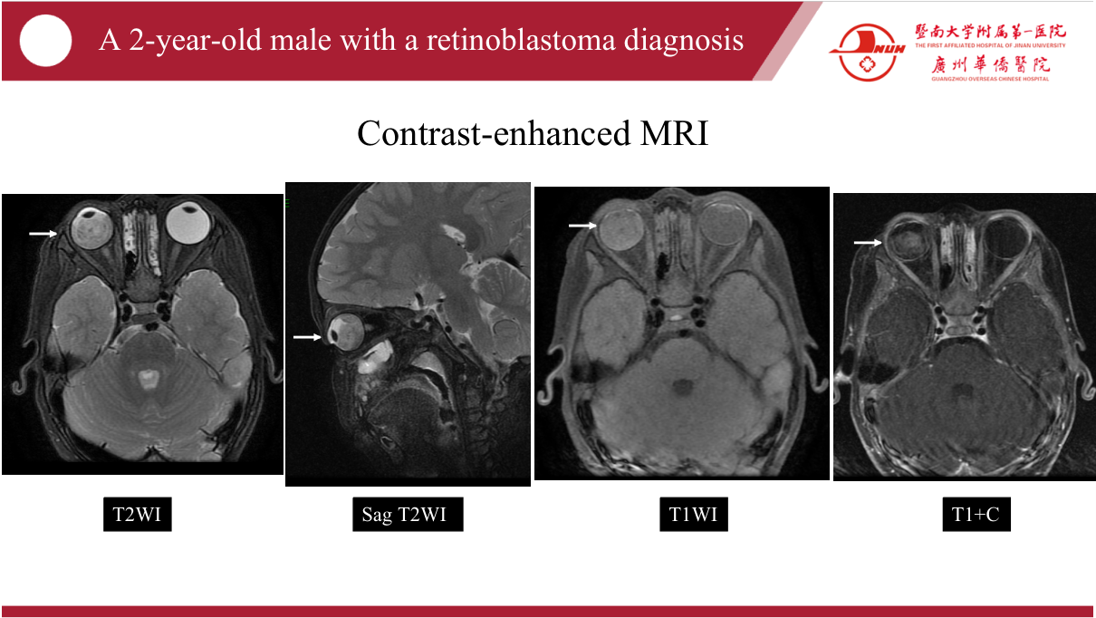
| MRI (with contrast) | Best for tumour extent, soft tissue, skull base/foramen involvement, intracranial extension, retropharyngeal nodes |
| CT | Bone erosion (skull base), calcification, staging (nodes, lungs) |
| PET-CT | Staging/metastasis, recurrence |
| Nasopharyngoscopy + biopsy | Diagnostic confirmation (histology) — EBV DNA levels for monitoring |
Imaging features: mass in the fossa of Rosenmüller / lateral pharyngeal recess; obliteration of pharyngeal recess, soft-tissue mass, skull base erosion, asymmetric nasopharyngeal soft tissue.
Staging/treatment: early — radiotherapy (NPC is very radiosensitive); locally advanced — chemoradiation; metastatic — palliative chemo. EBV DNA for surveillance.
2. Case II — the submandibular mass
Case: 54-year-old female, right submandibular mass 1 year, 1 cm, no hoarseness/dysphagia — the mass turned out to be related to nasopharyngeal carcinoma (cervical nodal metastasis).
The lesson: an apparently isolated neck mass can be a nodal metastasis from a hidden head & neck primary (NPC, thyroid, oral) — the workup must include nasopharynx, oral cavity, larynx, thyroid assessment — "always look for the primary."
Neck mass differential:
| Mass | Features |
|---|---|
| Submandibular gland tumour | Pleomorphic adenoma (most common salivary tumour — benign, slow); malignant (mucoepidermoid, adenoid cystic) |
| Lymph node metastasis | NPC, thyroid, oral SCC — hard, fixed |
| Lymphoma | Rubber, multiple levels |
| Thyroid (goitre/nodule) | Anterior midline, moves with swallowing |
| Branchial cyst / lymphangioma | Congenital, cystic |
| Infective (abscess/TB adenitis) | Painful/tender; TB nodes — caseous |
Imaging approach to a neck mass: US (superficial — thyroid/salivary/nodes) → MRI/CT with contrast (deep/extent) → FNA/core biopsy for tissue. If nodal metastasis found → search the primary: nasopharyngoscopy + panendoscopy + imaging of the whole upper aerodigestive tract.
3. Other high-yield head & neck topics (from the case discussion)
- Thyroid nodules: US TIRADS (hypoechoic, irregular margins, microcalcifications, taller-than-wide, high vascularity → suspicious) → FNA; calcitonin if medullary suspected.
- Parapharyngeal/retropharyngeal masses: location (pre vs post-styloid — parotid tail vs carotid body tumour), carotid body tumour = splaying of carotid bifurcation ("lyre sign").
- Sinus disease: sinonasal tumour vs chronic sinusitis (bone destruction, extension).
- Oral cavity SCC: tongue/mouth — MRI for depth of invasion + nodes; tobacco/alcohol risk.
High-Yield Points
| Topic | Must-remember |
|---|---|
| NPC endemic | Southern China/SE Asia — EBV-associated |
| NPC presentation | Unilateral glue ear (adult), epistaxis, cervical nodes, CN VI palsy |
| NPC best imaging | MRI (extent/soft tissue) + CT (bone erosion) |
| NPC diagnosis | Nasopharyngoscopy + biopsy; EBV DNA monitoring |
| NPC treatment | Radiotherapy (radiosensitive) ± chemo |
| Unilateral glue ear | Adult → NPC until proven otherwise |
| Neck mass + metastasis | Always search for hidden primary (NPC, thyroid, oral) |
| Salivary tumour | Pleomorphic adenoma most common (benign) |
| Thyroid nodule US | TIRADS — suspicious features → FNA |
| Carotid body tumour | "Lyre sign" — splaying of carotid bifurcation |
Topic Summary
The head & neck cases centre on NPC — the EBV-associated Southern-China cancer presenting with unilateral glue ear, epistaxis, cervical lymphadenopathy, and CN VI palsy; MRI (tumour extent) + CT (bone erosion) + nasopharyngoscopy/biopsy; treated with radiotherapy ± chemo. The second case teaches that a solitary neck mass may be a nodal metastasis from a hidden primary — always search the nasopharynx/oral cavity/thyroid. Neck mass workup: US → MRI/CT → FNA/biopsy; thyroid nodules by TIRADS → FNA; carotid body tumour by the lyre sign.
LMCHK OSCE Practice — Unilateral Glue Ear in an Adult
Station setup: A 45-year-old man presents with right-sided hearing loss and a blocked ear for 3 weeks; otoscopy shows right middle-ear effusion (glue ear). He has also noted blood-stained sputum and a lump in his neck for 2 months. He was born in Guangdong.
Candidate tasks (8 min):
- Recognise the red flag: adult unilateral glue ear = exclude NPC (especially with nasal/neck symptoms + Southern China origin).
- Take a focused history (epistaxis, nasal obstruction, headache, diplopia (CN VI), trismus, weight loss).
- Examine: cranial nerves (VI, V, III), cervical lymphadenopathy (posterior chain), nasopharyngoscopy (look for the mass).
- Order imaging: MRI (tumour extent, skull base, nodes) + CT (bone erosion) — and EBV serology/DNA.
- Discuss biopsy (nasopharyngoscopy-guided) for diagnosis and treatment (radiotherapy ± chemo); explain prognosis honestly.
Key marking cues:
- Unilateral glue ear in an adult → NPC workup — the exam pearl.
- Examines cranial nerves + nodes + nasopharynx.
- Orders MRI + CT + biopsy correctly.
- Knows the EBV association and treatment (RT ± chemo).