Preparatory Mindset
Cardiovascular imaging is the case-discussion chapter where plain-film cardiac anatomy + CT/MRI of the great vessels come together. The exam mindset: read the cardiac silhouette borders, recognise the classic chamber enlargements (mitral stenosis = left atrium; LV aneurysm = left border bulge), identify the acute aorta (dissection, aneurysm) and PE, and know the congenital shunt lesions (ASD/VSD — after the Y4S2 Pedi CHD chapter). The LMCHK angle: chest X-ray interpretation of the heart + CT aortogram for dissection + echo/CT for valves — the "which study for which cardiac question" competency.
Core Concepts
1. The cardiac silhouette on chest X-ray
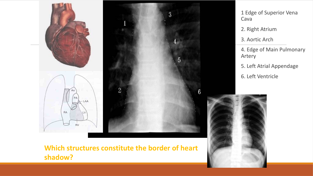
Which structures constitute the borders of the heart shadow?
| Border | Structure |
|---|---|
| Right border (upper → lower) | SVC / ascending aorta → right atrium |
| Left border (upper → lower) | Aortic knuckle → pulmonary trunk / left atrial appendage → left ventricle |
| Cardiophrenic angles | Right — IVC; left — fat pad |
| Double density | Enlarged left atrium (seen through the right heart border) |
Chamber enlargement patterns (exam table):
| Chamber | X-ray sign |
|---|---|
| Left atrium (LA) | Double density (right border), splaying of carina (>90°), left atrial appendage bulge (left border), posterior displacement (lateral film) — mitral stenosis/regurgitation |
| Left ventricle (LV) | Left border/bottom displacement ("LV contour" to the left and down) — HTN, aortic stenosis, LV failure |
| Right atrium (RA) | Right border prominence — ASD, tricuspid disease, RA enlargement |
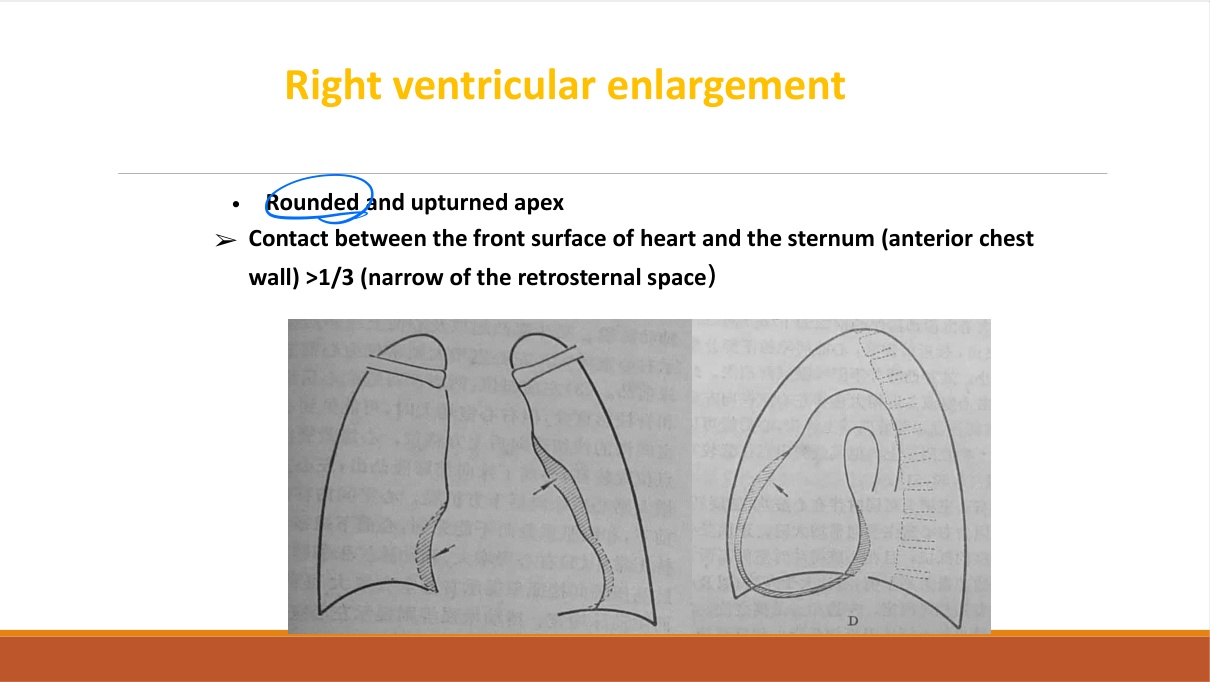
| Right ventricle (RV) | Upturned apex, filling of retrosternal space (lateral) — pulmonary HTN, cor pulmonale |
Heart size: cardiothoracic ratio >0.5 = cardiomegaly (PA film). Pericardial effusion = "water-bottle" enlarged globular heart (with clear lungs).
2. The acute aorta
| Condition | Imaging |
|---|---|
| Aortic dissection | CT aortogram — intimal flap separating true/false lumen; extent; arch vessel & pericardial involvement; Stanford A (ascending — surgical) vs B (descending — medical); TOE alternative; MRI if stable |
| Aortic aneurysm | CT/MRI sizing (>5.5 cm ascending, >5-5.5 cm abdominal → consider repair); mural thrombus; rupture = emergency (retroperitoneal haematoma) |
| Penetrating ulcer / intramural haematoma | CT — aortic wall abnormality without intimal flap |
3. Pulmonary embolism
- CTPA (CT pulmonary angiography) — the diagnostic test: filling defect (clot) in pulmonary artery; Westermark sign / Hampton hump on CXR (non-specific, historical).
- Clinical: sudden dyspnoea + pleuritic chest pain + tachycardia + hypoxaemia; Wells score → D-dimer → CTPA.
- Echo: McConnell sign (RV dysfunction) — supportive, not diagnostic.
4. Congenital heart disease (shunts) — imaging correlation
| Lesion | X-ray/CT findings |
|---|---|
| ASD | Cardiomegaly, prominent pulmonary trunk (pulmonary plethora), right atrial enlargement; fixed split S2 clinically |
| VSD | Cardiomegaly + pulmonary plethora; defect on echo/CT/MRI |
| PDA | Cardiomegaly + plethora; continuous murmur; calcified duct on CT |
| Coarctation | "3 sign" (aortic knob + post-stenotic dilatation), rib notching (inferior rib margins) |
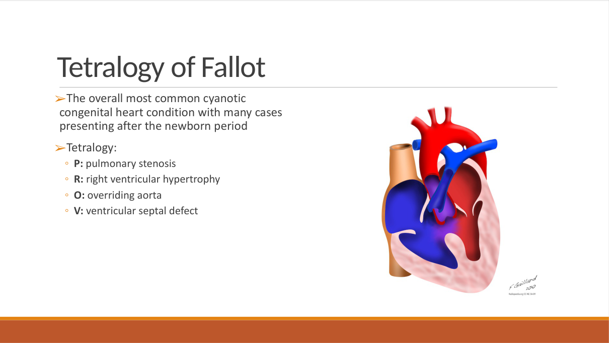
| Tetralogy of Fallot | "Boot-shaped" heart (upturned apex), right aortic arch (25%), pulmonary oligaemia |
5. Valvular and pericardial disease
| Disease | Imaging |
|---|---|
| Mitral stenosis (rheumatic) | LA enlargement (double density), splaying of carina, pulmonary venous hypertension/redistribution, Kerley B lines (chronic); echo = valve area/gradient |
| Aortic stenosis | LV hypertrophy/enlargement; calcified valve (CT/echo); pressure gradient on echo |
| Pericardial effusion | Globular "water-bottle" heart, clear lungs; echo confirms |
| Pericardial calcification (constrictive) | Curvilinear calcification (TB — China), small heart |
| Coronary artery disease | Coronary CTA (stenosis/plaque); calcium score (Agatston); myocardial perfusion; MRI viability (LGE) |
6. Cardiac MRI / echo — the "which study" pairs
- Echocardiography (TTE/TOE): first-line — valves, chambers, function (EF), pericardium, endocarditis vegetations.
- Cardiac MRI: myocardial viability (LGE = scar), cardiomyopathy phenotyping (HCM/amyloid), congenital anatomy.
- Coronary CTA: low-intermediate pre-test probability stable chest pain; anomalous coronaries.
- CAG (invasive coronary angiography): high-risk/ACS — intervention-ready.
High-Yield Points
| Topic | Must-remember |
|---|---|
| Right heart border | SVC/ascending aorta + RA |
| Left heart border | Aortic knuckle + pulmonary trunk + LA appendage + LV |
| LA enlargement | Double density + carina splaying >90° |
| RV enlargement | Upturned apex + retrosternal space filling |
| Cardiomegaly | CTR >0.5 |
| Pericardial effusion | Globular "water-bottle" heart, clear lungs |
| Dissection test | CT aortogram — intimal flap; type A = surgery |
| PE test | CTPA — filling defect |
| Coarctation | 3 sign + rib notching |
| TOF | Boot-shaped heart + pulmonary oligaemia |
| Mitral stenosis | LA enlargement + pulmonary redistribution + Kerley B |
| Coronary CTA | Stable chest pain, low-intermediate risk |
| Echo first-line | Valves, chambers, EF, pericardium |
Topic Summary
Cardiovascular imaging = cardiac silhouette reading (right/left borders, LA double density + carina splaying, RV upturned apex, CTR >0.5) + acute aorta (CT aortogram for dissection — flap, type A vs B) + CTPA for PE + congenital shunts (plethora vs oligaemia; coarctation 3-sign + rib notching; TOF boot-shaped heart) + valvular/pericardial disease (mitral stenosis LA enlargement + Kerley B; water-bottle pericardial effusion). Modality pairing: echo first-line; coronary CTA for stable chest pain; CAG for ACS; cardiac MRI for viability/cardiomyopathy.
LMCHK OSCE Practice — Cardiac CXR Interpretation
Station setup: A 62-year-old woman with rheumatic mitral stenosis presents with progressive dyspnoea. You are shown a PA chest X-ray.
Candidate tasks (8 min):
- Read the film systematically: cardiac size (CTR), borders, lung fields, hila.
- Identify the mitral stenosis pattern: LA enlargement (double density, carina splaying), pulmonary venous redistribution (upper lobe vessels prominent), Kerley B lines (interstitial oedema).
- Explain the haemodynamics: LA pressure ↑ → pulmonary venous congestion → interstitial oedema → dyspnoea.
- Recommend next investigation: echocardiography (valve area, gradient, LA size, PA pressure) — the definitive assessment.
- Discuss management direction (medical — diuretics, rate control; consider valvotomy/valve replacement if severe) and why imaging findings matter (progression to pulmonary hypertension).
Key marking cues:
- Systematic film-reading order (size, borders, lungs, hila).
- Recognises LA enlargement + redistribution + Kerley B = mitral stenosis pattern.
- Echo is the next step.
- Explains the haemodynamic link between X-ray and symptoms.