Preparatory Mindset
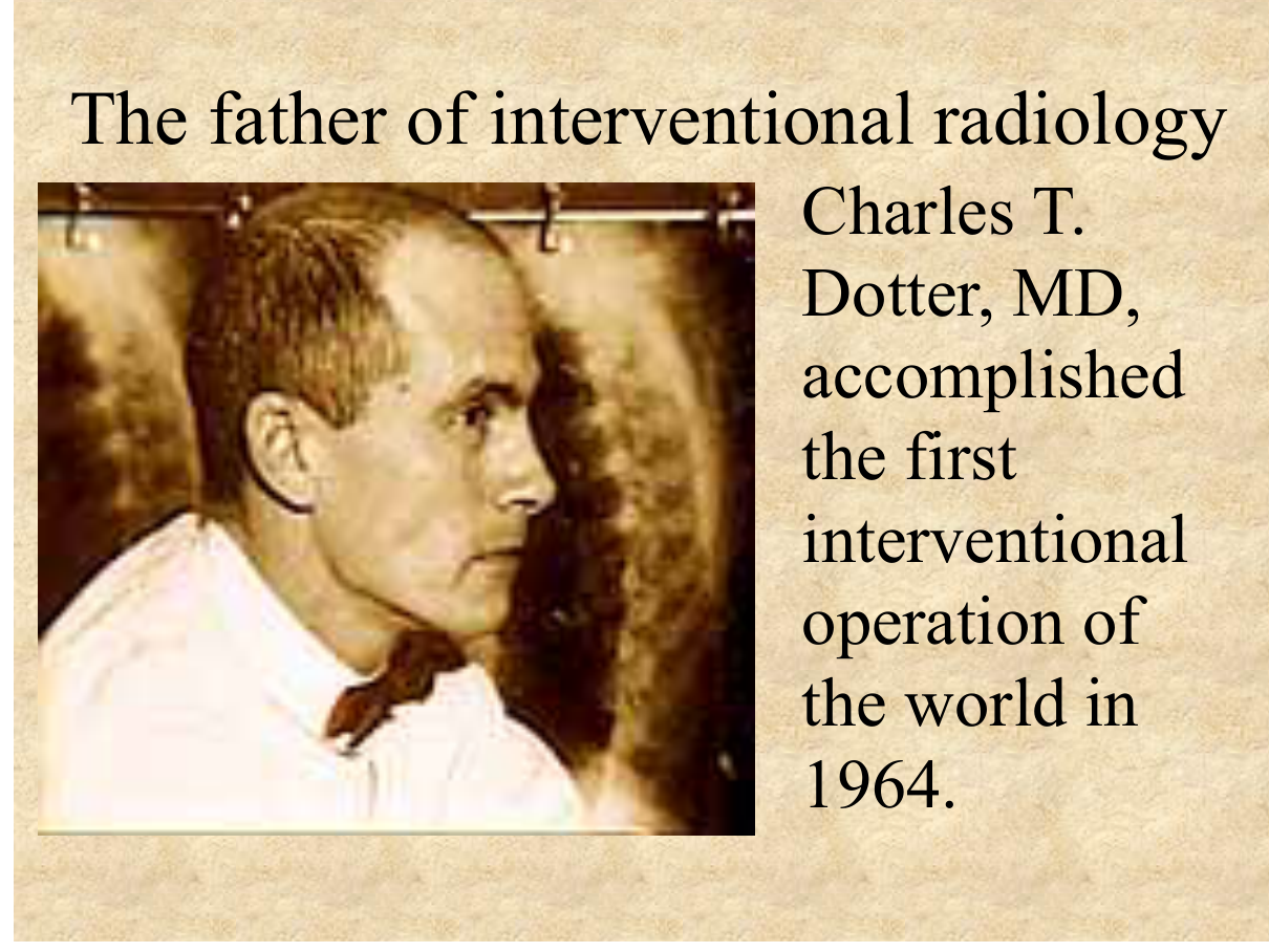
Interventional radiology (IR) is the "minimally invasive, image-guided treatment" chapter — the elective's IR III covers the principles and case discussions. The mindset: IR treats disease through needles, catheters, and wires under imaging guidance — the major categories: (1) embolisation (bleeding, tumours — TACE for HCC), (2) ablation (RFA/microwave for tumours), (3) biopsy/drainage (diagnostic + therapeutic), (4) vascular intervention (angioplasty, stents, thrombolysis, TIPS), (5) access (PICC, ports, dialysis catheters). The exam pearl: IR is the "organ-preserving" alternative to surgery — the father of IR (Charles Dotter, "father of interventional radiology") pioneered angioplasty in 1964. The LMCHK angle: know what IR can do for each clinical scenario (GI bleeding → embolisation, HCC → TACE, abscess → drainage, biliary obstruction → PTBD/stent).
Core Concepts
1. What is interventional radiology?
Definition: minimally invasive, image-guided diagnosis and treatment using needles, catheters, and wires — performed under fluoroscopy, CT, US, or MRI guidance.
The categories (the exam framework):
| Category | Procedures | Examples |
|---|---|---|
| Embolisation | Occlude a vessel | TACE (transarterial chemoembolisation — HCC), GI bleeding embolisation, PPH (uterine artery embolisation for fibroids), splenic/renal trauma bleeding, varicocele, AV malformation |
| Ablation | Destroy tissue in situ | RFA/microwave ablation of HCC/renal/lung tumours, bone metastases (pain), varicose veins (endovenous laser) |
| Biopsy / drainage | Tissue diagnosis / fluid evacuation | CT/US-guided biopsy (lung, liver, kidney, bone), abscess drainage (percutaneous catheter), pleural/ascitic drain, nephrostomy (obstructed kidney) |
| Vascular | Restore/redirect flow | Angioplasty + stenting (SFA, iliac, carotid, renal), thrombolysis (acute limb ischaemia, PE — catheter-directed), TIPS (portal hypertension), IVC filter |
| Access | Long-term vascular access | PICC, tunnelled central lines (ports), dialysis catheters |
2. The headliner cases
Case: HCC — TACE (transarterial chemoembolisation):
- Why: intermediate-stage (BCLC-B) multinodular HCC, not resectable/ablatable/transplantable.
- How: selective catheterisation of the hepatic artery (tumour is arterial-fed) → infusion of chemotherapy (doxorubicin/cisplatin) + embolic particles (or drug-eluting beads) → ischaemia + localised chemo.
- Imaging: pre-procedure CT/MRI (tumour burden, portal vein patency — TACE contraindicated with portal vein thrombosis unless segmental), CTA for arterial anatomy; post-procedure CT with lipiodol uptake (retained contrast in tumour = response marker).
- Complications: post-embolisation syndrome (fever, pain, nausea — common, self-limited), liver failure (poor reserve), abscess, cholecystitis.
Case: GI bleeding — embolisation:
- Indications: upper/lower GI bleed failing endoscopic control, unstable, recurrent.
- How: CT angiography (localise bleeding — extravasation) → selective angiography → embolisation (coils/gelfoam/particles) of the bleeding vessel.
- Advantages: avoids surgery, precise, repeatable; risk: bowel ischaemia (choose superselective embolisation).
Case: abscess — percutaneous drainage:
- How: US/CT-guided catheter insertion into the abscess → drainage + lavage; culture; antibiotics.
- When: liver abscess, intra-abdominal abscess, lung empyema, psoas abscess — often avoids surgery; large/multiloculated may need surgery.
Case: acute limb ischaemia / DVT — catheter-directed thrombolysis:
- Acute arterial occlusion (embolus/thrombus) → catheter-directed thrombolysis (tPA) ± aspiration thrombectomy; DVT (iliofemoral) → thrombolysis to prevent post-thrombotic syndrome.
3. The "which IR procedure" quick table (exam)
| Clinical scenario | IR procedure |
|---|---|
| HCC (intermediate, multinodular) | TACE |
| HCC (small, <3 cm, early) | RFA/MWA ablation (or surgery) |
| GI bleed (failed endoscopy) | Embolisation |
| Liver abscess | Percutaneous drainage |
| Obstructive jaundice (inoperable) | PTBD (percutaneous transhepatic biliary drainage) / biliary stent |
| Portal hypertension (variceal bleed, ascites) | TIPS |
| Fibroid (symptomatic, uterus-preserving) | UAE (uterine artery embolisation) |
| Renal artery stenosis (refractory HTN) | Angioplasty ± stent |
| Acute limb ischaemia | Catheter-directed thrombolysis ± thrombectomy |
| Suspicious lung nodule | CT-guided biopsy |
| Dialysis access | AV fistula creation / tunnelled catheter / fistula angioplasty |
High-Yield Points
| Topic | Must-remember |
|---|---|
| IR definition | Image-guided minimally invasive treatment (needles/catheters/wires) |
| Father of IR | Charles Dotter (1964, angioplasty) |
| TACE | HCC (BCLC-B) — chemo + embolisation of hepatic artery; contraindicated with portal vein thrombosis (unless segmental) |
| Post-embolisation syndrome | Fever, pain, nausea — common, self-limited |
| GI bleed IR | Embolisation after failed endoscopy; CTA localises first |
| Abscess | Percutaneous drainage + antibiotics |
| RFA | Small HCC (<3 cm) — ablation |
| TIPS | Portal hypertension (varices, ascites) |
| UAE | Fibroids — uterus-preserving |
| PTBD | Obstructive jaundice, inoperable |
| Acute limb ischaemia | Catheter-directed thrombolysis ± thrombectomy |
| IR advantages | Organ-preserving, less invasive, faster recovery, repeatable |
Topic Summary
Interventional radiology is image-guided minimally invasive treatment — the five pillars: embolisation (TACE for HCC — chemoembolic to the arterial-fed tumour; GI bleeding embolisation), ablation (RFA for small HCC), biopsy/drainage (CT/US-guided; abscess drainage often avoids surgery), vascular (angioplasty/stents, catheter-directed thrombolysis, TIPS), and access (PICC/ports/dialysis catheters). The IR "which procedure" map (TACE for BCLC-B HCC, RFA for small HCC, embolisation for bleeding, drainage for abscess, TIPS for portal hypertension, UAE for fibroids) is the exam core. IR's value: organ-preserving, less invasive, faster recovery, repeatable — the modern alternative to surgery.
LMCHK OSCE Practice — Referral for IR (HCC)
Station setup: A 60-year-old man with HBV cirrhosis is found to have multinodular HCC (three lesions: 4 cm, 3 cm, 2 cm) with preserved liver function (Child-Pugh A), patent portal vein, no extrahepatic metastasis. He is not a candidate for resection or transplant.
Candidate tasks (8 min):
- Stage: BCLC intermediate (B) — multinodular, preserved liver function, no portal invasion.
- Recommend TACE (transarterial chemoembolisation) — the standard for BCLC-B.
- Explain the procedure (hepatic artery catheterisation → chemo + embolisation; HCC is arterial-fed), pre-procedure imaging (CT/MRI + CTA for anatomy), and why portal vein patency matters (TACE contraindicated with main portal vein thrombosis — embolic risk to liver).
- Discuss post-embolisation syndrome (fever, pain — self-limited) and follow-up (CT with lipiodol uptake as response marker, re-TACE as needed).
- Mention alternatives: ablation for the small lesions (RFA/MWA), systemic therapy if progression, and liver transplant candidacy review.
Key marking cues:
- BCLC staging → TACE for intermediate HCC.
- Knows portal vein patency requirement for TACE.
- Explains arterial feeding of HCC + embolisation concept.
- Covers post-embolisation syndrome + imaging follow-up.
- Presents treatment alternatives objectively.