Preparatory Mindset
CNS imaging is the case-discussion chapter for brain tumours, haemorrhage, and ischaemia — the imaging workhorse of neurology. The mindset: CT first for the acute brain (haemorrhage vs ischaemia), MRI for tumour characterisation (DWI/ADC, enhancement, location). The exam focus: the tumour differential by location and age (high-grade glioma vs low-grade pilocytic; meningioma — extra-axial, dural; pituitary — sellar; metastasis — multiple, ring-enhancing), the haemorrhage patterns (SAH — berry aneurysm, epidural vs subdural, hypertensive ICH, amyloid), acute stroke imaging (NCCT to exclude bleed → CTPA/MRI-DWI for ischaemia → thrombolysis window). The LMCHK angle: "CT brain — name the haemorrhage/tumour" and the acute stroke decision path.
Core Concepts
1. Brain tumours — the case-driven differential
| Tumour | Location / age | Imaging features |
|---|---|---|
| High-grade glioma (GBM) | Supratentorial, adult | Irregular enhancing mass, necrosis, oedema (butterfly — corpus callosum), heterogeneous; DWI restricted |
| Low-grade glioma (pilocytic astrocytoma) | Posterior fossa / cerebellum, child/young adult | Well-defined cyst + enhancing mural nodule (pilocytic — the "cyst with nodule") |
| Meningioma | Extra-axial (dural-based), convexity/parasagittal, adult (F>M) | Homogeneous enhancing extra-axial mass, dural tail sign, calcification, hyperostosis; well-defined |
| Pituitary adenoma | Sella turcica | Sellar/suprasellar mass, enlargement of sella, cavernous sinus extension; haemorrhage (pituitary apoplexy) |
| Metastasis | Multiple, grey-white junction, adult (lung, breast, melanoma) | Multiple ring-enhancing lesions + surrounding vasogenic oedema |
| Schwannoma (CPA) | Cerebellopontine angle (CN VIII — acoustic neuroma) | IAC widening, enhancing CPA mass |
| Epidermoid / arachnoid cyst | CPA/suprasellar | DWI restriction (epidermoid) vs follows CSF (arachnoid) |
| Craniopharyngioma | Suprasellar, child | Calcified cystic suprasellar mass |
| Lymphoma | Periventricular, immunocompromised | Homogeneous enhancing, restricted DWI |
MRI sequences to remember: T1 (anatomy), T2/FLAIR (oedema — white), DWI (ischaemia/tumour cellularity — restricted = bright), ADC (dark when restricted), T1 + contrast (enhancement = BBB breakdown), GRE/SWI (haemorrhage/calcification), MRS (elevated choline = tumour), PWI (perfusion).
2. Intracranial haemorrhage — patterns
| Type | Location | Cause | Imaging |
|---|---|---|---|
| SAH (subarachnoid) | Basal cisterns, Sylvian fissure | Berry aneurysm rupture (most common), trauma | CT — hyperdense blood in cisterns ("star sign"); CT angiogram for aneurysm; LP if CT negative (xanthochromia) |
| Epidural haematoma (EDH) | Biconvex/lens-shaped, does not cross sutures | Middle meningeal artery tear (temporal fracture) | Biconvex hyperdense mass; "lucid interval" clinically |
| Subdural haematoma (SDH) | Crescent-shaped, crosses sutures | Bridging vein tear (elderly, anticoagulation, shaken baby) | Crescent over convexity; chronic — isodense/hypodense |
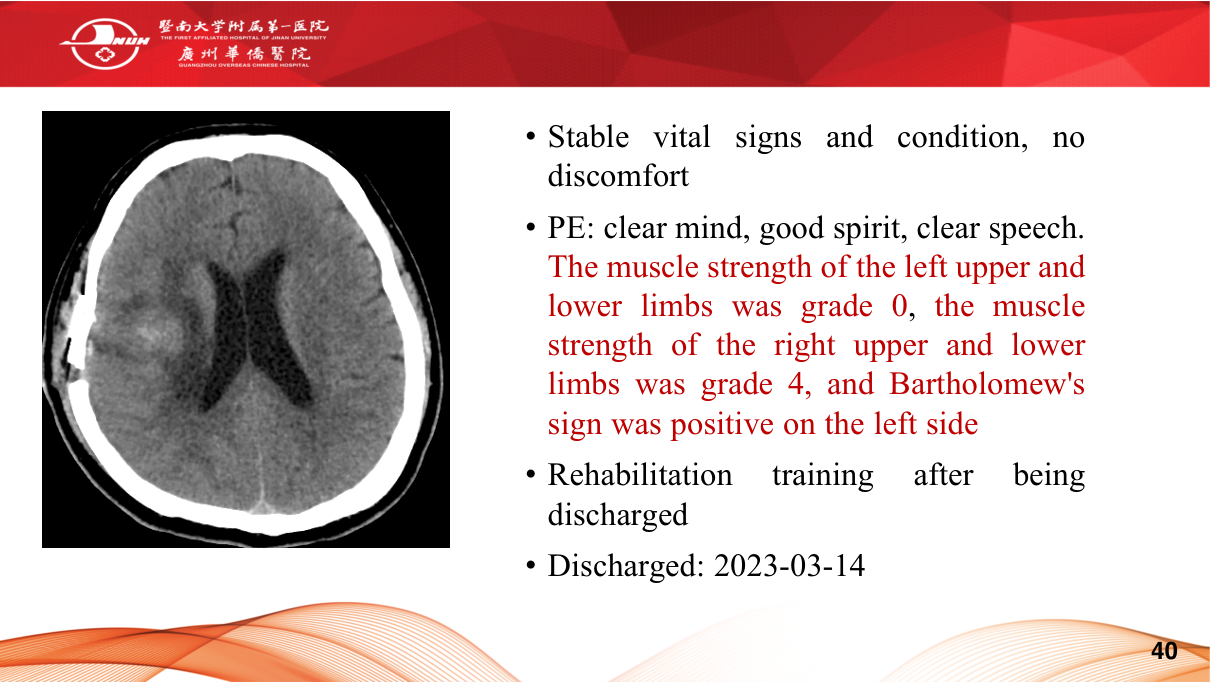
| Intracerebral (ICH) | Basal ganglia, thalami, lobes | Hypertension (basal ganglia — most common), amyloid (lobar, elderly), anticoagulation | Hyperdense parenchymal haematoma ± oedema, mass effect |
| Intraventricular | Ventricles | Extension of ICH, aneurysm | Hyperdense in ventricles |
| Cerebellar haemorrhage | Posterior fossa | HTN | Surgical emergency if >3 cm (brainstem compression) |
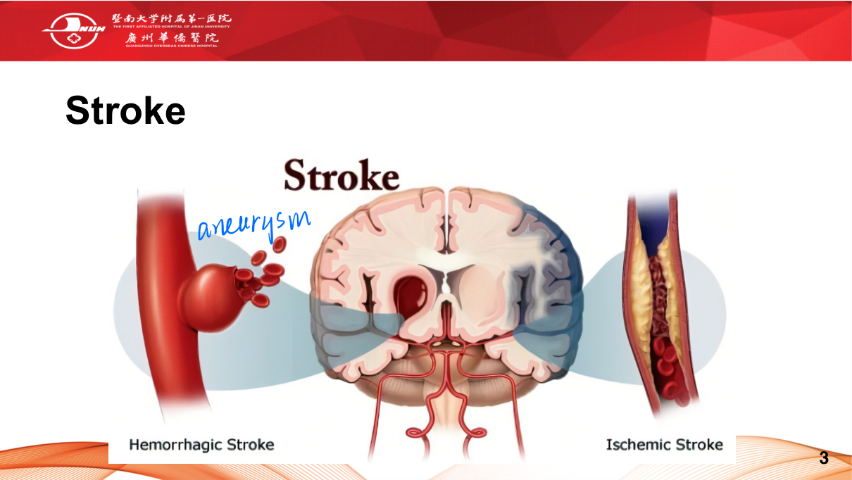
3. Acute ischaemic stroke
The imaging pathway:
- NCCT first — exclude haemorrhage (the thrombolysis gatekeeper); early signs: loss of grey-white differentiation, insular ribbon sign, dense MCA sign (thrombus), sulcal effacement.
- MRI-DWI — hyperintense (restricted diffusion) within minutes — the most sensitive for acute ischaemia; ADC dark.
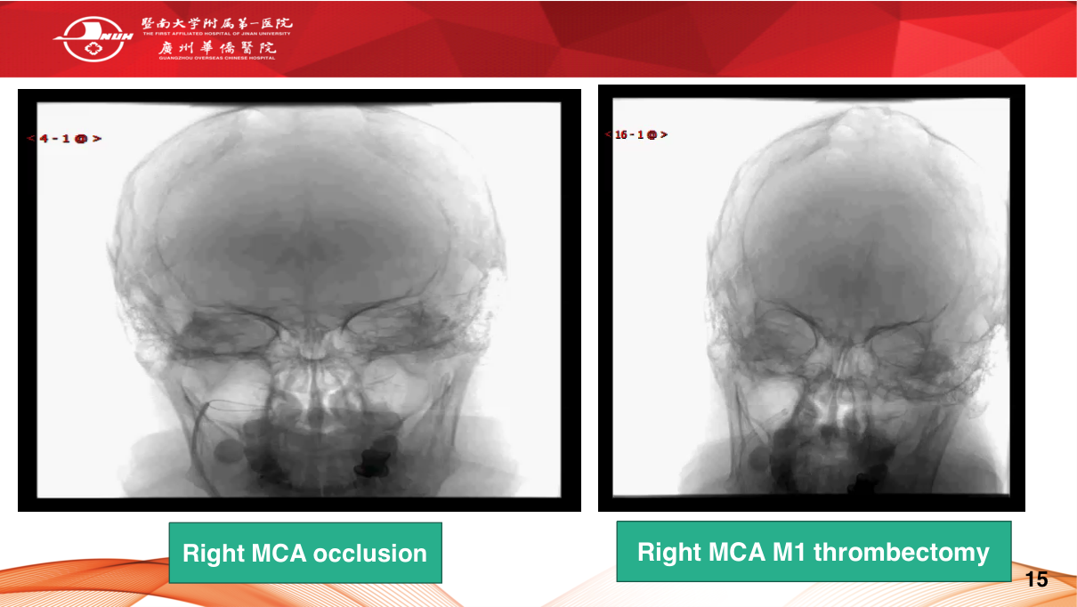
- CTA — large vessel occlusion (LVO — thrombectomy candidate); CTP/MRI PWI — penumbra (ischaemic but salvageable tissue — MTT ↑, CBV normal) vs core (CBV ↓).
- Decision: thrombolysis (alteplase) within 4.5 h, thrombectomy within 6-24 h (LVO + salvageable penumbra).
Territories: MCA (most common — contralateral hemiparesis + hemisensory loss + hemianopia + aphasia if dominant); ACA (leg > arm); PCA (hemianopia); brainstem/cerebellar (vertebrobasilar — vertigo, ataxia, cranial nerves).
4. Other high-yield cases
- Cerebral abscess: ring-enhancing lesion with thin smooth rim, restricted DWI (pus), satellite lesions — fever + immunocompromise/otogenic.
- Toxoplasmosis vs lymphoma (HIV): toxo — multiple ring-enhancing basal ganglia; lymphoma — periventricular, homogeneous, restricted DWI.
- Tuberculoma / TB meningitis: basal meningeal enhancement, hydrocephalus, tuberculomas.
- Multiple sclerosis: periventricular ovoid "Dawson fingers", DWI negative, enhancing (active), dissemination in space/time.
- Hydrocephalus: ventricular dilatation (obstructive vs communicating), periventricular oedema (transependymal flow).
- Venous sinus thrombosis: empty delta sign on contrast CT, thrombus on MRI/MRV — headache, seizures, risk (OCP, dehydration).
High-Yield Points
| Topic | Must-remember |
|---|---|
| Acute brain first test | NCCT (exclude haemorrhage) |
| Acute ischaemia | MRI-DWI bright / ADC dark — most sensitive |
| Stroke window | Thrombolysis ≤4.5 h; thrombectomy ≤6-24 h (LVO + penumbra) |
| Dense MCA sign | Thrombus in MCA on NCCT |
| High-grade glioma | Necrosis + oedema + enhancement, adult supratentorial |
| Pilocytic astrocytoma | Cyst + enhancing mural nodule (child, cerebellum) |
| Meningioma | Extra-axial, dural tail, homogeneous enhancement |
| Metastasis | Multiple ring-enhancing + vasogenic oedema |
| SAH | CT — cisternal blood; CTA for berry aneurysm; LP if CT negative |
| EDH | Biconvex, lens-shaped, doesn't cross sutures (MMA tear) |
| SDH | Crescent, crosses sutures (bridging vein, elderly/anticoagulation) |
| HTN ICH | Basal ganglia most common |
| Abscess | Ring-enhancing + restricted DWI (pus) |
| Empty delta sign | Venous sinus thrombosis |
Topic Summary
CNS imaging is CT-first (acute) and MRI (characterisation). Brain tumours are sorted by location, age, and enhancement — high-grade glioma (necrosis/oedema, adult), pilocytic astrocytoma (cyst + nodule, child), meningioma (extra-axial, dural tail), metastasis (multiple ring-enhancing), pituitary (sellar). Haemorrhage patterns: SAH (cisterns + aneurysm), EDH (biconvex, doesn't cross sutures), SDH (crescent, crosses sutures), hypertensive ICH (basal ganglia). Acute stroke: NCCT (bleed) → MRI-DWI (ischaemia, bright within minutes) → CTA/CTP (LVO + penumbra) → thrombolysis ≤4.5 h / thrombectomy ≤24 h. Abscess (ring + restricted DWI), MS (Dawson fingers), venous thrombosis (empty delta) complete the case set.
LMCHK OSCE Practice — Acute Stroke Decision
Station setup: A 68-year-old man presents with right-sided weakness and aphasia for 90 minutes. BP 160/90, glucose 6.0. NCCT brain shows no haemorrhage, but loss of grey-white differentiation in the left MCA territory and a dense left MCA sign.
Candidate tasks (8 min):
- Recognise acute left MCA ischaemic stroke (right hemiparesis + aphasia = dominant hemisphere) — within the window.
- Interpret NCCT: no haemorrhage (thrombolysis eligible); early ischaemic signs (loss of grey-white, dense MCA = thrombus).
- State contraindications to thrombolysis (recent surgery/bleeding, anticoagulation, BP >185/110 uncontrolled, glucose extremes, prior ICH) and order CTA/CTP (LVO → thrombectomy; penumbra assessment).
- Plan: alteplase (IV thrombolysis) within 4.5 h (start immediately after eligibility confirmed) + thrombectomy if LVO with salvageable penumbra (≤24 h).
- Admit to stroke unit, swallow assessment, BP management (not too low acutely), monitoring.
Key marking cues:
- NCCT is the gatekeeper (bleed excluded → thrombolysis).
- Interprets early ischaemic signs (dense MCA, loss of grey-white).
- Knows thrombolysis window (4.5 h) + contraindications.
- Escalates to CTA/thrombectomy for LVO.
- Mentions stroke unit + swallowing + BP.