Preparatory Mindset
Ultrasound is the first-line, real-time, radiation-free imaging modality — the elective lecture ("Advancement and Case Discussion of Ultrasonography") covers contrast-enhanced ultrasound (CEUS), 3D/4D ultrasound, elastography, and Doppler. The exam mindset: US answers "is it cystic or solid, vascular or not, hard or soft" in real time, at the bedside, with no radiation — ideal for focal liver lesions (CEUS), thyroid/breast nodules, fetal assessment (3D/4D), and vascular cases (Doppler). CEUS is the standout exam topic: microbubble contrast agents are strictly intravascular — enhancement patterns characterise focal liver lesions (hemangioma = peripheral nodular → centripetal fill; HCC = arterial hyperenhancement + washout). Elastography adds tissue stiffness (cirrhosis staging, thyroid/malignancy). The LMCHK angle: US is the first-line test for RUQ pain, pelvic/obstetric assessment, and superficial masses.
Core Concepts
1. Ultrasound basics
- Principles: high-frequency sound waves (2-15 MHz); reflections from tissue interfaces form the image; no ionising radiation; real-time; portable.
- Transducers: convex (abdomen), linear (superficial — thyroid, breast, vessels), sector/phased (cardiac), endocavitary (transvaginal, transrectal).
- Structures imaged: abdomen (liver, gallbladder, kidney, pancreas, spleen), neck, breast, eyes, pelvis, vessels, thyroid, scrotum.
- Grey-scale: anechoic (fluid — black), hypoechoic, isoechoic, hyperechoic (calcification/gas — white), posterior acoustic enhancement (cyst), shadowing (stone/calcification).
2. Contrast-Enhanced Ultrasound (CEUS)
- Microbubble contrast agents (e.g., SonoVue — sulphur hexafluoride) — strictly intravascular (do not leak into interstitium) — perfect for real-time vascular assessment.
- Applications: focal liver lesion characterisation (the classic), cardiac (contrast echo), carotid plaque, renal, prostate, sentinel nodes.
- Liver lesion enhancement patterns (the exam table):
| Lesion | Arterial phase | Portal/venous phase |
|---|---|---|
| Hemangioma | Peripheral nodular enhancement → centripetal fill | Persistent fill ("pooling") — stays bright |
| HCC | Arterial hyperenhancement (early, intense) | Washout (becomes hypoechoic) |
| FNH | Arterial hyperenhancement + spoke-wheel / stellate vascularity | Iso/hyperechoic (central scar) |
| Metastasis | Rim/irregular enhancement | Washout (early) |
| Hepatocellular adenoma | Arterial enhancement | Iso/washout (variable) |
3. Elastography
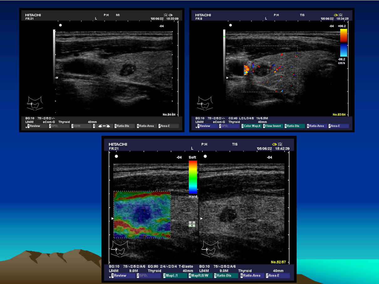
- Principle: measures tissue stiffness (shear-wave / strain) — malignant lesions are stiffer.
- Uses: liver fibrosis staging (non-invasive alternative to biopsy — kPa correlates with fibrosis stage), thyroid nodule malignancy risk, breast lesions, lymph nodes.
- Pearl: stiff thyroid nodule + suspicious US features → FNA; stiff liver → advanced fibrosis.
4. 3D / 4D ultrasound
- 3D: volume acquisition → multiplanar reconstruction (coronal/sagittal/axial) — fetal anomaly assessment, surface rendering of the fetal face.
- 4D = real-time 3D — live volume imaging (fetal movement, cardiac).
- Uses: fetal malformations (cleft lip, neural tube), pelvic pathology, breast volume.
5. Doppler ultrasound
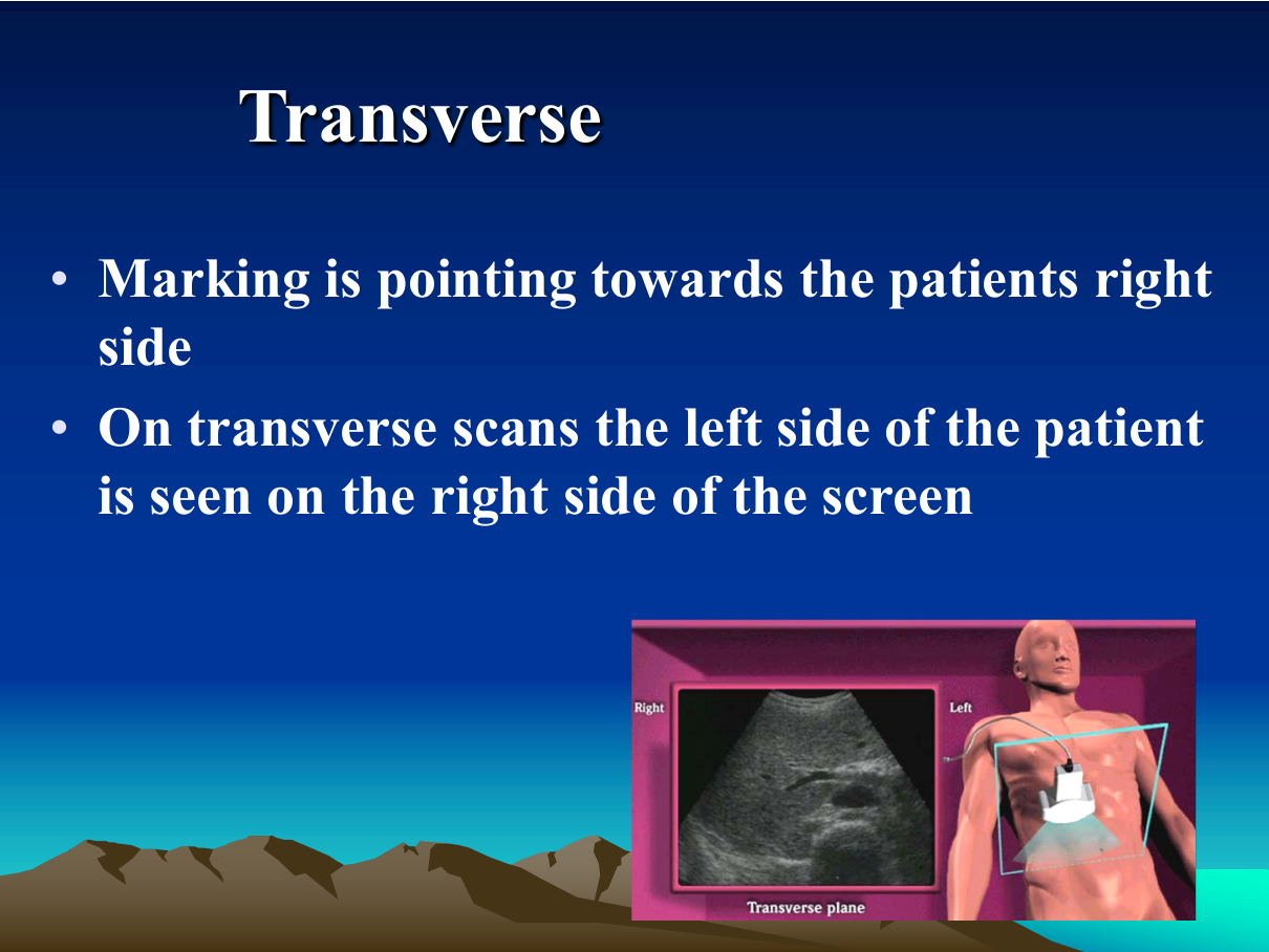
- Colour Doppler: flow direction/velocity mapping (red = toward, blue = away).
- Spectral Doppler: velocity waveform analysis (PSV, EDV, RI).
- Uses: carotid stenosis (PSV), DVT (venous compressibility + flow), renal artery stenosis, hepatic vasculature (portal HTN, Budd-Chiari), scrotal torsion (absent flow — emergency), obstetrics (umbilical artery Doppler — AEDF/REDF = fetal compromise), varicocele, AV fistula.
6. Case themes (from the lecture)
- Focal liver lesion — CEUS characterisation (the headliner).
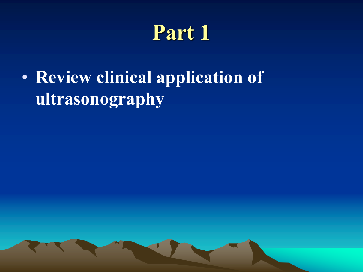
- Thyroid nodule — US features (hypoechoic, irregular margins, microcalcifications, taller-than-wide) + elastography → TIRADS → FNA.
- Breast mass — US BI-RADS (cyst vs solid, margins), Doppler vascularity.
- Fetal US — 3D/4D surface rendering, anomaly screening.
- Vascular — carotid, DVT, portal hypertension.
High-Yield Points
| Topic | Must-remember |
|---|---|
| US advantages | Real-time, no radiation, bedside, low cost |
| CEUS contrast | Microbubbles — strictly intravascular |
| Hemangioma CEUS | Peripheral nodular → centripetal fill, persists |
| HCC CEUS | Arterial hyperenhancement + washout |
| FNH CEUS | Spoke-wheel/stellate arterial + central scar |
| Elastography | Stiffness — liver fibrosis staging, thyroid/malignancy |
| 3D/4D | Fetal anomaly assessment, surface rendering |
| Doppler | Carotid PSV, DVT, scrotal torsion (emergency), umbilical AEDF |
| Cyst US | Anechoic + posterior enhancement |
| Stone US | Hyperechoic + shadowing |
Topic Summary
Ultrasound is the real-time, radiation-free first-line modality. CEUS (microbubble, strictly intravascular) characterises focal liver lesions by enhancement pattern — hemangioma (peripheral nodular → fill, persists), HCC (arterial hyperenhancement + washout), FNH (spoke-wheel). Elastography measures stiffness (liver fibrosis, thyroid/malignancy); 3D/4D US serves fetal anomaly assessment; Doppler assesses vascular flow (carotid, DVT, torsion — emergency, umbilical AEDF). The LMCHK-relevant skill: choose US first for superficial/abdominal/pelvic questions and interpret the classic enhancement patterns.
LMCHK OSCE Practice — RUQ Pain: the Focal Liver Lesion
Station setup: A 45-year-old woman has an incidental 3-cm liver lesion on US. The lesion is hyperechoic and well-defined. You are asked how to characterise it.
Candidate tasks (8 min):
- State the differential for a well-defined hyperechoic liver lesion: hemangioma (most common), FNH, HCC, metastasis, adenoma.
- Recommend the best next step: CEUS or MRI with contrast (MRI is definitive — hemangioma is pathognomonic).
- Describe the CEUS pattern of hemangioma (peripheral nodular enhancement → centripetal fill → persistent bright on delayed phase) vs HCC (arterial hyperenhancement + washout).
- Mention elastography role (stiffness) and Doppler (flow).
- Explain that biopsy is usually NOT needed for a typical hemangioma (risk of bleeding) — imaging diagnosis suffices.
Key marking cues:
- Knows the hyperechoic liver lesion differential.
- CEUS/MRI enhancement patterns — the core competency.
- Avoids unnecessary biopsy of hemangioma.
- Links imaging to clinical context (known cirrhosis → HCC risk).