Preparatory Mindset
Musculoskeletal (MSK) imaging covers trauma (fractures) and bone tumours — two parts in the elective. The mindset for fractures: describe by location, pattern, displacement/angulation, alignment, and joint involvement; recognise the classic named fractures (Colles, scaphoid, femoral neck, supracondylar, ankle). For bone tumours: age and location are the two most important discriminators — osteosarcoma (adolescent, metaphysis of long bones — "sunburst"), Ewing sarcoma (child/young adult, diaphysis — "onion peel"), giant cell tumour (20-40, epiphysis — "soap bubble"), chondrosarcoma (adult, axial/pelvis), metastasis (elderly, multiple, lytic/blastic), osteoid osteoma (child, night pain relieved by NSAIDs — nidus). The LMCHK angle: "describe this fracture" and "child with bone pain — benign vs malignant lesion."
Core Concepts
1. Fracture — systematic description
- Location — bone, site (proximal/midshaft/distal), intra-articular?
- Pattern — transverse, oblique, spiral (twisting — non-accidental injury in children), comminuted, greenstick (child), buckle (torus — child, compression).
- Displacement/angulation — % of shaft width, direction, shortening.
- Alignment — joint congruity.
- Soft tissues — joint effusion (fat pad sign — elbow), open fracture (air in soft tissues).
Fracture types by mechanism:
| Type | Feature | Typical |
|---|---|---|
| Transverse | Perpendicular | Direct blow |
| Oblique/spiral | Angled/spiral | Twisting — spiral = child abuse red flag |
| Comminuted | Multiple fragments | High energy |
| Greenstick | Incomplete (one cortex) — child | Bending force |
| Torus (buckle) | Buckling of cortex — child | Compression |
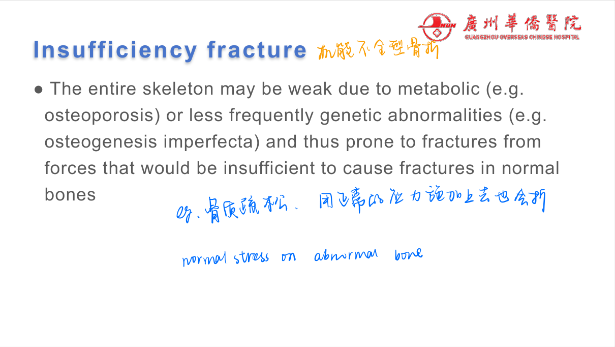
| Pathological | Through abnormal bone (tumour, cyst, osteoporosis) | Minimal trauma |
| Stress (fatigue/insufficiency) | Fatigue — abnormal stress normal bone (runners); insufficiency — normal stress abnormal bone (osteoporosis) | Repetitive load |
2. Classic named fractures (high-yield)
| Fracture | Site | Features |
|---|---|---|
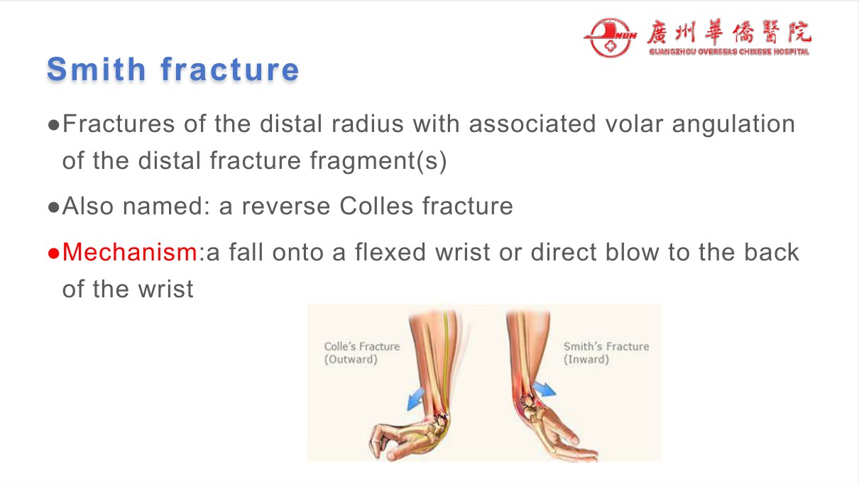
| Colles | Distal radius | Dinner-fork deformity (dorsal displacement + angulation), fall on outstretched hand (FOOSH); ulnar styloid often |
| Smith | Distal radius | Reverse (volar displacement) |
| Scaphoid | Carpal scaphoid | FOOSH; snuffbox tenderness; avascular necrosis risk (proximal pole); occult on initial X-ray — repeat/CT/MRI |
| Femoral neck / intertrochanteric | Proximal femur | Elderly fall; unable to weight-bear; NOF fracture vs intertrochanteric (surgical fixation) |
| Supracondylar humerus | Distal humerus (child) | Elbow fall; fat pad sign; neurovascular injury (brachial artery, median nerve) — check pulses |
| Ankle (Weber) | Malleoli | Weber A (below syndesmosis), B (at), C (above — unstable) |
| Clavicle | Midshaft | FOOSH/shoulder; most common fracture in children |
| Talus/calcaneus | Foot | Fall from height (calcaneus — also lumbar spine) |
| Hip dislocation | Femoral head | Posterior — shortened internally rotated leg |
Child abuse (NAI) — must-know red flags: spiral/humeral/femoral fractures in non-walking child, multiple fractures at different healing stages, metaphyseal corner fractures, posterior rib fractures, subdural haemorrhage, retinal haemorrhage — skeletal survey + safeguarding referral.
3. Bone tumours — age + location = diagnosis
| Tumour | Age | Location | Imaging |
|---|---|---|---|
| Osteosarcoma | 10-25 (adolescent) | Metaphysis of long bones (distal femur, proximal tibia) | "Sunburst" (spiculated periosteal reaction), Codman triangle, soft-tissue mass; malignant |
| Ewing sarcoma | 5-20 | Diaphysis (femur, tibia, pelvis) | "Onion-peel" periosteal reaction, permeative lytic, large soft-tissue mass; malignant |
| Giant cell tumour | 20-40 | Epiphysis (distal femur, proximal tibia, distal radius) | Eccentric "soap-bubble" lytic, extends to subchondral bone; locally aggressive (benign) |
| Osteochondroma | Child/adolescent | Metaphysis (cartilage-capped exostosis) | Most common benign — pedunculated/sessile outgrowth; malignant transformation rare |
| Osteoid osteoma | Child/young adult | Diaphysis (femur, tibia) | Small lucent nidus + sclerosis; night pain relieved by NSAIDs |
| Enchondroma | Adult | Phalanges/metaphysis | Lytic with "ring-and-arc" calcification; benign |
| Chondrosarcoma | >40 | Axial/pelvis, proximal femur | Lobulated, ring-and-arc calcification, cortical destruction — malignant |
| Metastasis | Elderly | Multiple, axial (spine, pelvis, proximal femur) | Lytic (lung, renal, thyroid, myeloma), blastic (prostate, breast), pathological fracture |
| Multiple myeloma | Elderly | Skull, spine, ribs | "Punched-out" lytic lesions, diffuse osteopenia; M protein |
| Aneurysmal bone cyst (ABC) | Child/adolescent | Metaphysis (posterior elements) | Expansile "blown-out" cystic, fluid-fluid levels (MRI) |
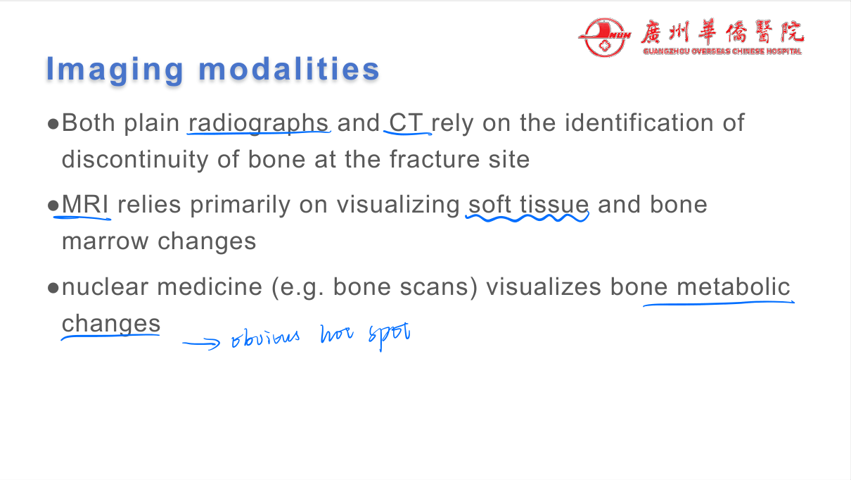
Bone tumour workup: plain film (first — matrix/mineralisation, periosteal reaction) → MRI (marrow + soft tissue extent) → CT (matrix/cortex) → biopsy (staging — chest CT, bone scan/PET).
Benign vs malignant — the descriptors:
| Feature | Benign | Malignant |
|---|---|---|
| Margins | Well-defined, sclerotic rim | Ill-defined, permeative |
| Periosteal reaction | Solid/absent | Sunburst/onion-peel/Codman (interrupted) |
| Cortex | Intact/expanded | Destroyed |
| Soft-tissue mass | Absent | Present |
| Growth | Slow/stable | Progressive |
High-Yield Points
| Topic | Must-remember |
|---|---|
| Fracture description | Location, pattern, displacement, alignment, joint, soft tissue |
| Greenstick/torus | Childhood fractures (incomplete/buckle) |
| Spiral fracture + child | NAI red flag |
| Colles | Dinner-fork, dorsal displacement, FOOSH |
| Scaphoid | Snuffbox pain; AVN risk; occult initially |
| Femoral neck | Elderly fall — surgical |
| Supracondylar | Fat pad sign; brachial artery/median nerve risk |
| Osteosarcoma | Adolescent, metaphysis, sunburst |
| Ewing | Child, diaphysis, onion-peel |
| Giant cell | 20-40, epiphysis, soap bubble |
| Osteoid osteoma | Night pain + NSAIDs relief, nidus |
| Metastasis | Elderly, multiple; lytic vs blastic (prostate) |
| Myeloma | Punched-out lytic, M protein |
| Malignant features | Ill-defined margins, interrupted periosteal reaction, cortical destruction, soft-tissue mass |
Topic Summary
Fractures are described systematically (location, pattern, displacement, alignment) — the named fractures (Colles dinner-fork, scaphoid AVN risk, femoral neck, supracondylar fat-pad + neurovascular risk) and childhood patterns (greenstick, torus; spiral = NAI red flag) are the exam staples. Bone tumours are diagnosed by age + location: osteosarcoma (adolescent metaphysis, sunburst), Ewing (child diaphysis, onion-peel), giant cell (20-40 epiphysis, soap bubble), chondrosarcoma (adult axial), metastasis (elderly, multiple); benign lesions have well-defined margins + solid periosteal reaction, malignant have permeative margins + interrupted periosteal reaction + cortical destruction + soft-tissue mass. Workup: plain film → MRI → biopsy.
LMCHK OSCE Practice — The Painful Bone Lesion
Station setup: A 14-year-old boy presents with 2 months of left distal thigh pain and swelling, worse at night, with a palpable mass. X-ray shows a permeative lytic lesion in the distal femoral metaphysis with "sunburst" periosteal reaction, Codman triangle, and a soft-tissue mass.
Candidate tasks (8 min):
- Recognise osteosarcoma — adolescent + distal femoral metaphysis + sunburst periosteal reaction + soft-tissue mass.
- Explain the imaging features (sunburst = malignant periosteal reaction; Codman triangle = interrupted periosteum; soft-tissue mass = aggressive).
- Staging workup: MRI (marrow + soft-tissue extent), CT chest (lung metastasis — the most common site), bone scan/PET, biopsy (orthopaedic oncology) — pre-biopsy staging.
- Discuss treatment: neoadjuvant chemotherapy → wide local excision (limb salvage) → adjuvant chemo; amputation if not salvageable.
- Counsel on prognosis (cure ~60-70% with modern multimodal therapy, better if no metastasis).
Key marking cues:
- Age + location + sunburst = osteosarcoma — the classic triad.
- Describes malignant features correctly.
- Stages before biopsy (chest CT for lung mets).
- Knows neoadjuvant chemo + surgery treatment sequence.
- Gives honest prognosis.