# Ch04: Chest Radiology — Specific Disorders(胸部疾病影像诊断)
Preparatory Mindset
This chapter covers the specific chest disorders from the teacher's Part III lecture: pneumonia (bacterial, viral/mycoplasma, fungal, immunocompromised), lung abscess, pulmonary tuberculosis, sarcoidosis, diffuse interstitial fibrosis, radiation pneumonitis, collagen vascular diseases, pneumoconiosis (coal workers', asbestos), and airway diseases (COPD/emphysema, bronchiectasis). The major purpose of CXR in suspected chest infection is to establish whether pneumonia is present — identifying the responsible organism radiologically is rarely possible because appearances overlap.
Core Concepts
Bacterial pneumonia
- One or more areas of consolidation, from a small ill-defined opacity to a whole lobe.
- Cavitation may occur within consolidated areas.
- DDx: pulmonary edema, pulmonary infarction.
- Common CAP lobar pneumonia organism: Streptococcus pneumoniae.
- Lung abscess (complication): localized suppurative lesion; spherical opacity with central lucency ± air-fluid level; smooth inner wall, even wall thickness. Difficult to distinguish from cavitating neoplasm or Wegener's granulomatosis.
Viral and mycoplasma pneumonia
- Loss of clarity of vascular markings; widespread or localized ill-defined patchy consolidation.
- Pleural effusions are rare.
- The radiological abnormality may persist for many weeks after clinical recovery.
Pulmonary tuberculosis
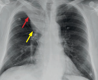
- Primary TB (first infection, usually childhood): Ghon focus = area of consolidation, usually mid/upper zones; primary complex = small pulmonary opacity (sometimes invisible) + enlarged hilar/mediastinal lymph nodes. Heals and often calcifies (calcified primary complex remains visible for life).
- Spread: bronchial tree → tuberculous bronchopneumonia (patchy/lobar, often multi-lobar, bilateral, frequently cavitates); bloodstream → miliary TB (innumerable small nodules, uniform in size/density/distribution).
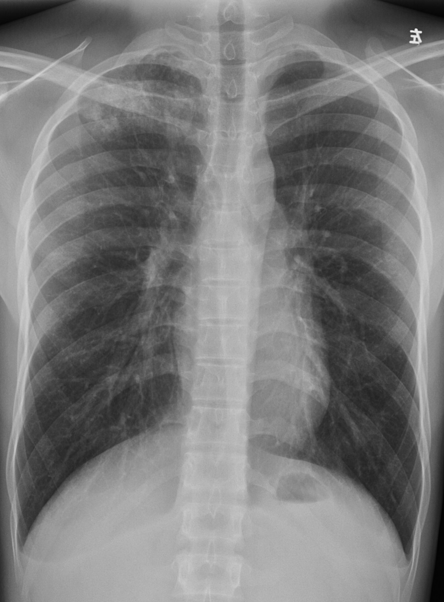
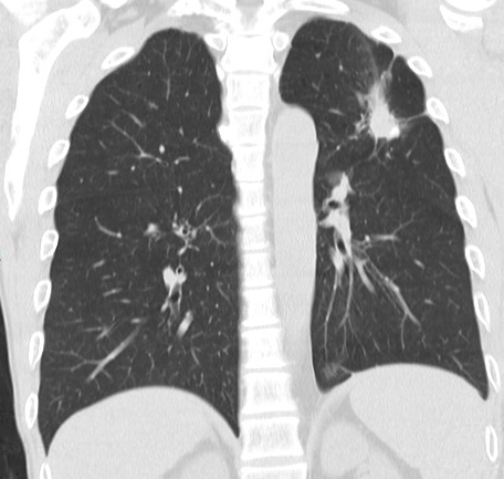
- Secondary (reactivation) TB: cough, hemoptysis, weight loss, night sweats, malaise. Location: apical and posterior segments of upper lobes; apical segments of lower lobes. Ill-defined consolidations; cavitation common.
- Progression: consolidation → cavitation → fibrosis ± calcification; pleural effusions leave permanent thickening/calcification.
- Is TB active? Active: ill-defined opacities, cavitation, new lesions on serial films, positive sputum. Stable/inactive: well-defined opacities, more calcification, lack of change over time.
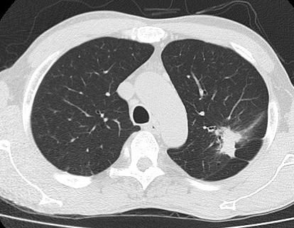
- Key signs: cavity with smooth inner wall, spherical opacities, tree-in-bud sign.
Fungal pneumonia
- Normal host (histoplasmosis, coccidioidomycosis, blastomycosis): similar to TB, cavitation a particular feature; healing by fibrosis and calcification.
- Aspergillosis (Aspergillus fumigatus) three ways: ① colonize a pre-existing cavity → fungus ball/mycetoma; ② pneumonia in the immunocompromised; ③ allergic bronchopulmonary aspergillosis.
Pneumonia in the immunocompromised host
- More susceptible to infection; atypical radiographic appearance; usually widespread non-specific opacification (may be indistinguishable from edema, hemorrhage or neoplastic disease).
- Opportunistic fungi, TB, Pneumocystis jirovecii (in AIDS: widespread low-density air-space opacity, ground-glass opacification, reticular opacities/septal thickening).
Sarcoidosis
- Non-caseating granulomas in many organs; diagnosis by correlating clinical, pathological and radiological features.
- Chest findings: bilateral and symmetrical hilar and paratracheal lymphadenopathy; reticulonodular opacities maximal in mid and upper zones → pulmonary fibrosis (irreversible). ~10% develop significant lung involvement.
Diffuse interstitial pulmonary fibrosis
- Known causes: extrinsic allergic alveolitis, collagen vascular diseases (notably rheumatoid arthritis), drug-induced fibrosis, pneumoconiosis, sarcoidosis, idiopathic (UIP/IPF/cryptogenic fibrosing alveolitis).
- UIP: thickening of alveolar walls with fibrosis; progression → breakdown of alveolar walls → small rounded air-spaces → 'honeycomb lung' (restrictive defect, severe reduction in gas transfer). Imaging: hazy basal opacities → loss of vessel clarity → honeycombing (ill-defined nodules with connecting lines & circular lucencies, subpleural, basal predominance) → pulmonary hypertension → cardiomegaly. Higher incidence of lung cancer in these patients.
Radiation pneumonitis
- After radiotherapy for intrathoracic neoplasms and breast carcinoma.
- Initially no change → a few weeks later ill-defined small opacities in the radiation field → fibrosis with dense coarse opacification.
Collagen vascular diseases
- Wegener's granulomatosis: one or more well-defined consolidations/masses, usually mid zones, which may cavitate; difficult to distinguish from bronchogenic carcinoma (single) or metastases (multiple).
Pneumoconiosis
- Coal workers' pneumoconiosis: many small nodules similar to miliary TB → progressive massive fibrosis (homogeneous rounded opacities in the upper halves of the lungs).
- Asbestos-related disease: ① pleural plaques (minor exposure) — localized, some calcified, harmless, indicate prior exposure; ② diffuse pleural thickening (may encase lungs → restrictive); ③ asbestosis — bilateral, maximal at bases; ④ malignant mesothelioma and bronchial carcinoma at far higher frequency.
Airway diseases
- Chronic bronchitis = clinical diagnosis (productive cough ≥3 consecutive months in 2 successive years); CXR usually normal; if abnormal → complication (emphysema, pneumonia, cor pulmonale).
- Emphysema = increase beyond normal size of air-spaces distal to the terminal bronchiole with destructive wall changes. Imaging: increased lung volume (low flat diaphragm — midpoints below 7th rib anteriorly/12th rib posteriorly; elongated narrow heart; wide intercostal spaces); attenuated vessels (reduced size/number; bulla = sharply demarcated focal region).
- Bronchiectasis = irreversible dilatation of bronchi, often with impaired drainage → persistent infection. HRCT diagnostic: signet ring sign; CXR: tram-track opacities. Causes: recurrent infection, cystic fibrosis, ciliary dyskinesia.
High-Yield Points
- Key Point: Bacterial pneumonia = consolidation + air bronchogram; viral/mycoplasma = patchy, ill-defined, no effusion.
- Key Point: Primary TB = Ghon complex (Ghon focus + hilar nodes); secondary TB = upper lobe apical/posterior cavitary disease; miliary = uniform small nodules.
- Key Point: Active TB = ill-defined opacities + cavitation + new lesions; healing = calcification + well-defined margins.
- Key Point: Sarcoidosis = bilateral symmetrical hilar adenopathy + mid/upper zone reticulonodular opacities.
- Key Point: UIP/IPF = subpleural basal honeycombing.
- Key Point: Emphysema = hyperinflation (low flat diaphragm, wide rib spaces) + attenuated vessels + bullae.
- Key Point: Asbestos → pleural plaques (benign) but also mesothelioma and bronchial carcinoma.
- Key Point: Bronchiectasis = signet ring sign on HRCT.
LMCHK OSCE Practice(OSCE & LMCHK)
- "Small nodule + bilateral hilar adenopathy" → sarcoidosis (stage I); with mid/upper reticulonodular opacities → stage II.
- "Cavitating upper lobe lesion in a young patient with hemoptysis + night sweats" → reactivation TB; describe cavity + tree-in-bud + apical location.
- "Mycetoma" = fungus ball in a pre-existing cavity (aspergilloma), changes position with patient posture.
- Pneumonia in AIDS with bilateral ground-glass → Pneumocystis jirovecii.
- Remember the rule: CXR rarely identifies the causative organism of pneumonia — the job is to confirm pneumonia is present.
Topic Summary
Specific chest disorders cluster into infections (bacterial/viral/mycoplasma/fungal/TB/immunocompromised), granulomatous disease (sarcoidosis), interstitial disease (UIP/IPF, radiation, collagen vascular), occupational disease (coal, asbestos) and airway disease (COPD/emphysema, bronchiectasis). Master the classic signatures: lobar consolidation + air bronchogram (S. pneumoniae), upper-lobe cavitary disease + tree-in-bud (reactivation TB), bilateral hilar adenopathy (sarcoidosis), subpleural honeycombing (UIP), hyperinflation + bullae (emphysema), signet ring sign (bronchiectasis).
Illustrations(图解速览)
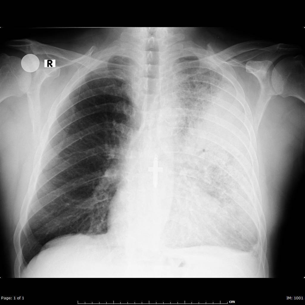
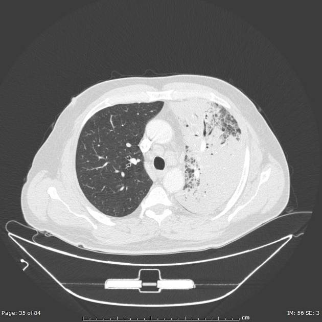
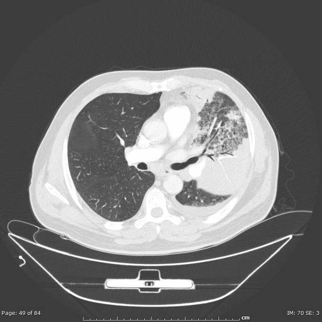
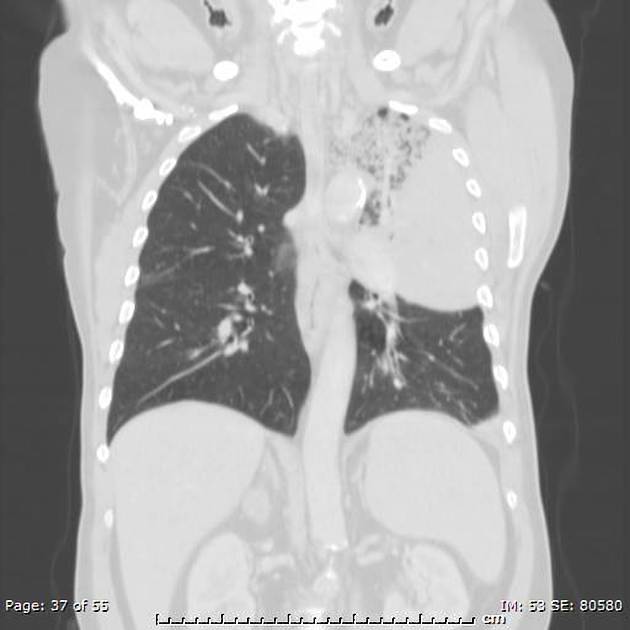
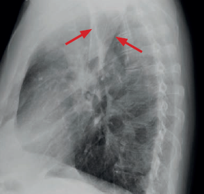
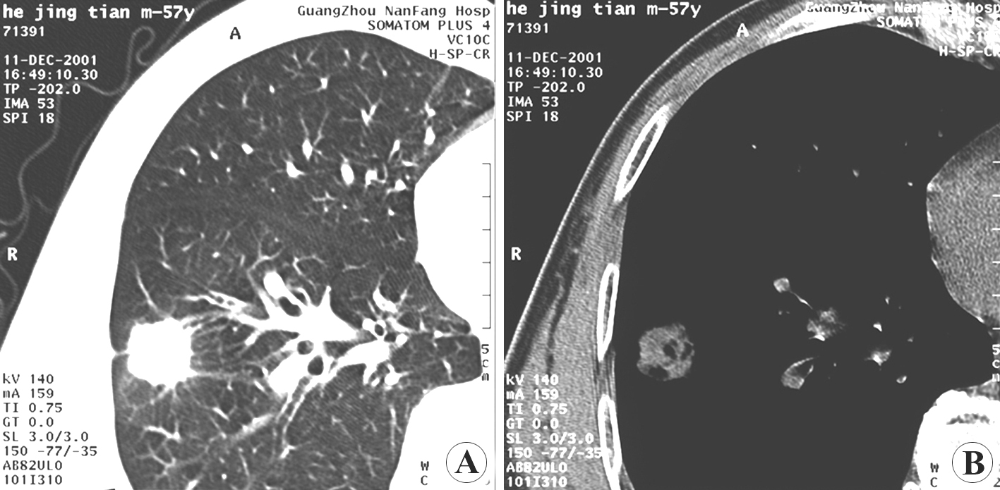
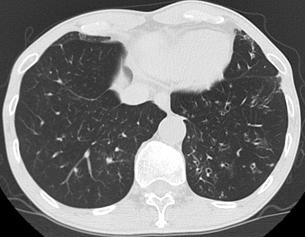
Case gallery for this chapter — identify the imaging technique, location, features and diagnosis for each: