# Ch18: Skeletal Trauma Imaging(骨创伤影像)
Preparatory Mindset
Skeletal trauma imaging: plain radiographs diagnose the presence and type of a fracture; CT gives better assessment and soft-tissue extent (and is often safer for the severely injured); MRI shows a dark line across the marrow fat with increased signal from hemorrhage/edema; radionuclide bone scanning shows increased activity within 2–3 days, persisting while fractures heal. Look for direct signs (fracture line, step in the cortex, interruption of trabeculae, buckling of the cortex) and indirect signs (soft tissue swelling, joint effusion).
Core Concepts
Types of fracture
- Comminuted (粉碎性)
- Transverse / oblique / spiral (横贯/斜行/螺旋)
- Impacted (嵌入)
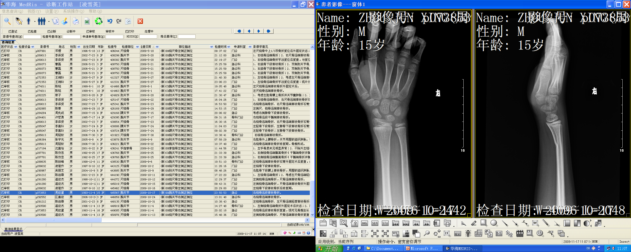
- Greenstick (青枝) — children, incomplete
- Avulsion (撕裂)
- Burst / crush (爆裂/压缩)
- Insufficiency (不全)
Radiographic signs of fracture (direct & indirect)
- Fracture line (lucent or dense); step in the cortex; interruption of bony trabeculae; bulging/buckling of the cortex; soft tissue swelling; joint effusion.
Dislocation
- Joint surfaces no longer maintain their normal relationship; stress views demonstrate ligamentous rupture (e.g. inversion stress opens the lateral ankle joint = lateral ligament rupture).
Further plain film views
- Oblique view (radial head), flexion view (C2 arch), delayed films (occult fractures).
Salter–Harris classification (pediatric growth plate fractures)
- Type I: epiphyseal separation through the growth plate.
- Type II (commonest): fragment of the metaphysis accompanies the displaced epiphysis.
- Type III: fracture through the epiphysis and growth plate.
- Type IV: fracture through epiphysis, growth plate and metaphysis.
- Type V: crush injury of the growth plate; usually no radiographic changes.
Regional injuries
- Clavicle fracture (upward displacement of medial fragment); acromioclavicular dislocation.
- Humeral neck fracture (elderly; may involve greater tuberosity); anterior shoulder dislocation (may fracture greater tuberosity); posterior shoulder dislocation (electric shock/epilepsy; "lightbulb" appearance of humeral head).
- Elbow dislocation: backward and lateral displacement of radius and ulna.
- Supracondylar fractures (children): potentially dangerous — brachial artery and nerve injury; epicondylar fractures before epiphyses fuse.
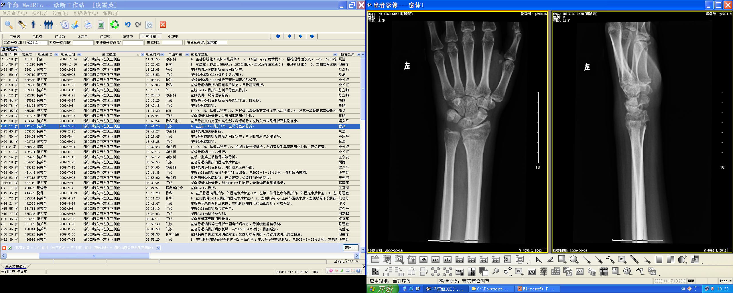
- Colles' fracture: fracture through the lower end of the radius; posterior displacement/angulation = "dinner fork" deformity; sometimes ulnar styloid avulsed; common in the elderly.
- Smith fracture: reverse deformity to Colles' — anterior displacement/angulation.
- Scaphoid fracture: across the waist; if missed → non-union or avascular necrosis; triquetral flake fracture only on lateral view.
- Carpal dislocation: lunate/capitate dislocate posterior to the lower radius (ligament rupture).
- Bennett's fracture: fracture through the base of the thumb metacarpal involving the carpometacarpal articular surface; requires internal fixation. Boxer's fracture: shaft of the little finger metacarpal, angulated.
- Pelvic fractures: pubic rami fractures, pubic symphysis separation.
- Femoral neck fracture: interrupts Shenton's line (imaginary line along the inferior border of the superior pubic ramus and the inferomedial border of the femoral neck — should be continuous and smooth; interruption = fractured neck of femur or developmental dysplasia of the hip DDH); impacted fracture disrupts trabecular architecture; intertrochanteric fracture between greater and lesser trochanters.
- Patellar fracture/dislocation.
- Knee: fat in haemarthrosis; tibial plateau (bumper) fracture.
- Ankle fractures: lateral/medial/both malleoli ± talus disruption in the ankle mortice; lateral shift of talus = ligamentous damage.
- Talar neck fracture.
- Calcaneal fracture: jumping injuries → compression/flattening; Bohler's angle <20°; CT shows subtalar joint disruption.
- Lisfranc's (tarsometatarsal) dislocation: lateral dislocation of metatarsals; step between medial cuneiform and base of 2nd metatarsal.
Stress, insufficiency and pathological fractures
- Stress fracture: repeated minor trauma (march fracture); sclerosis adjacent to the fracture line; MRI (STIR bright edema) and bone scan positive when radiographs normal.
- Insufficiency fracture: normal activity/minimal trauma in weakened bone (osteoporosis, osteomalacia); commonest = vertebral compression fractures; also sacrum, pubic rami, femoral necks.
- Pathological fracture: through a lesion (lytic metastases from breast cancer, myeloma, Paget's disease).
- Non-accidental injury (child abuse): multiple fractures at different healing stages, posterior rib fractures, metaphyseal corner fractures — must be recognized.
High-Yield Points
- Key Point: Direct signs: fracture line, step in cortex, interrupted trabeculae, cortical buckling; indirect: soft tissue swelling, joint effusion.
- Key Point: Salter-Harris: I through plate, II (commonest) metaphyseal fragment, III epiphysis+plate, IV epiphysis+plate+metaphysis, V crush.
- Key Point: Colles' = dinner-fork (posterior displacement); Smith = reverse (anterior).
- Key Point: Scaphoid waist fracture → risk of AVN/non-union if missed.
- Key Point: Shenton's line interruption = femoral neck fracture or DDH.
- Key Point: Bohler's angle <20° = calcaneal compression fracture.
- Key Point: Stress fracture: bone scan/MRI positive before plain films; insufficiency = weakened bone (osteoporosis).
- Key Point: Greenstick = children (incomplete); comminuted/burst = high energy.
- Key Point: Posterior shoulder dislocation = lightbulb sign (electric shock/epilepsy).
LMCHK OSCE Practice(OSCE & LMCHK)
- Hip x-rays, ankle x-ray (fibula fracture), Colles' fracture, femoral shaft tumour are listed LMCHK orthopedics stations. Be ready to describe: "Colles' fracture — distal radius fracture with posterior angulation (dinner-fork deformity)"; "femoral neck fracture — Shenton's line interrupted, impacted"; "ankle — lateral malleolus fracture with talar shift = unstable."
- Pediatric ortho (LMCHK): clubfoot, scoliosis, DDH, pediatric fracture, septic arthritis — DDH uses US (infant) + Shenton's line (older); pediatric fractures include greenstick and Salter-Harris.
- Osteoporotic (intertrochanteric) fracture in the elderly: hip x-ray; recognize in the LMCHK "intertrochanteric fracture (osteoporotic fracture)" common case.
- Non-accidental injury: multiple healing-stage fractures → safeguarding concern (child protection).
Topic Summary
Skeletal trauma: plain films first (direct + indirect signs), CT for complex/extent, MRI for occult (stress, marrow edema), bone scan for occult/stress. Know fracture types (greenstick, comminuted, avulsion, burst, insufficiency), the Salter-Harris classification, and regional signatures: Colles' (dinner-fork) vs Smith, scaphoid waist (AVN risk), Bennett's/boxer's, Shenton's line (femoral neck/DDH), Bohler's angle (calcaneus), Lisfranc's dislocation. Stress/insufficiency/pathological fractures and non-accidental injury complete the chapter.
Illustrations(图解速览)
Case gallery for this chapter — identify the imaging technique, location, features and diagnosis for each: