# Ch14: Neuroimaging — Brain Tumors, Stroke & Hemorrhage(颅脑疾病影像)
Preparatory Mindset
Brain disease imaging splits into tumors, vascular disease (infarction, hemorrhage, aneurysm/AVM), demyelination, degeneration, infection and trauma. The first question for any intracranial mass: is it intra-axial (within the brain — metastasis or astrocytoma in 75% of adult cases) or extra-axial (outside the brain — meningioma or schwannoma)? Extra-axial lesions push the brain away: look for CSF cleft, displaced subarachnoid vessels, gray matter between lesion and white matter, and a widened subarachnoid space.
Core Concepts
Intra-axial vs extra-axial
- Extra-axial signs: CSF cleft between lesion and brain; subarachnoid vessels displaced; gray matter between lesion and white matter; subarachnoid space widened (lesion pushes brain away). Meningioma has a broad dural base / dural tail of enhancement and adjacent bone hyperostosis.
- Intra-axial: arises within brain parenchyma (astrocytoma/GBM, metastasis, oligodendroglioma).
Meningioma (extra-axial, most common non-glial intracranial tumor)
- Arises from meninges of the vault, falx or tentorium; commonest sites: parasagittal, cerebral convexities, sphenoid ridges.
- WHO: Grade I typical/benign 88–94%; Grade II atypical 5–7%; Grade III anaplastic 1–2%.
- CT: hyperdense mass, calcification; homogenous enhancement on CECT; adjacent bone hyperostosis.
- MRI: iso- or slightly hypointense on T1WI and T2WI; broad-based dural attachment; dural tail sign; homogenous enhancing globose mass.
Glioma / astrocytoma (intra-axial)
- Glioma = non-specific term for tumors from glial cells (astrocytes — astrocytoma is the commonest).
- Low-grade astrocytoma: hyperintense on T2, no or mild enhancement; imaging may be subtle.
- Glioblastoma multiforme (GBM) — most common primary CNS malignancy: large size, crosses the midline, ill-defined outer margin, strong enhancement with irregular central necrosis (cauliflower pattern), peripheral white matter edema; subcortical white matter location.
- Oligodendroglioma: like low-grade astrocytoma but always contains calcification; mild hyperintensity T1, hyperintensity T2, no/mild enhancement.
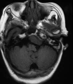
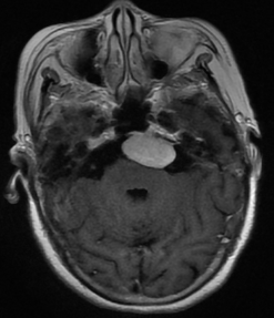
Vestibular schwannoma (acoustic neuroma) (extra-axial)
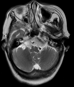
- Arises on the nerve within the internal auditory canal, extends medially into the cerebellopontine angle (CPA).
- 80% of CPA masses; solid nodular with cystic degeneration; enlargement of the ipsilateral internal auditory canal.
Pituitary tumors
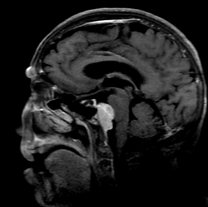
- Macroadenoma (>1 cm) vs microadenoma (<1 cm).
- Macroadenoma extends superiorly; indentation at the diaphragm sella → snowman appearance; compresses the optic chiasm; cavernous sinus invasion.
- Microadenoma: ~2/3 hypoattenuating on dynamic contrast-enhanced CT/MRI (enhances less than normal gland, best seen 30–60 min after contrast); patients present with hyperprolactinemia.
Brain metastases
- 25–30% of intracranial tumors in hospital patients.
- 80% from 5 primaries: lung, renal cell, breast, melanoma, GI adenocarcinomas (mostly colorectal).
- 80% localize to cerebral hemispheres, 15% cerebellum, 3% basal ganglia; often at the gray-white matter junction.
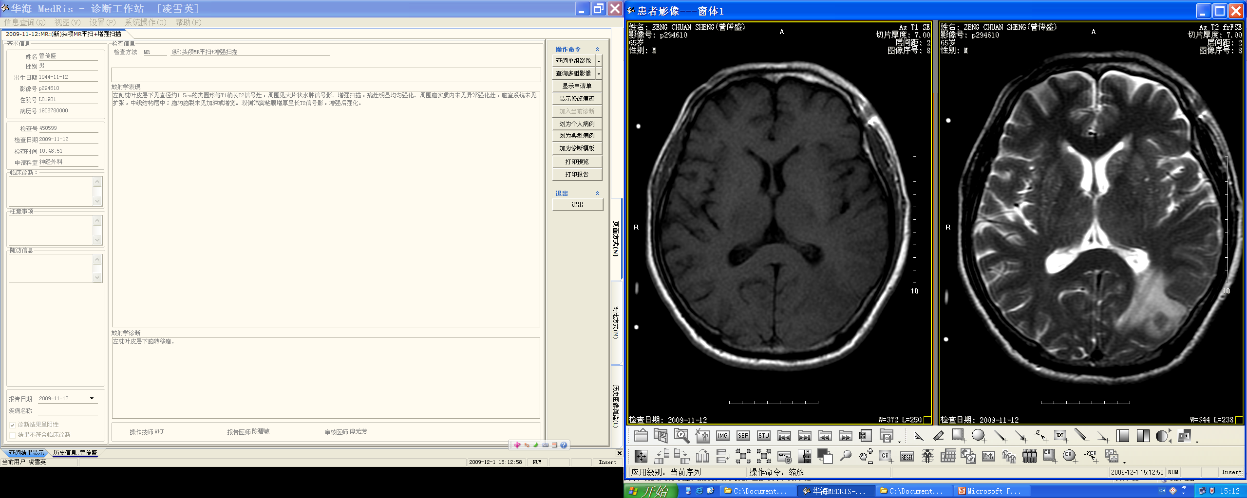
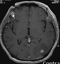
- "Small nodule, large edema" sign; typically sphere and well circumscribed (vs GBM's complex shape); rim-enhancing with necrotic center; abscess shows central restricted diffusion (DWI).
- Metastasis vs GBM: metastasis at gray-white junction, sphere, well circumscribed, less deep brain involvement; GBM in subcortical white matter, complex shape, extends to ependymal surface.
Cerebral hemorrhage
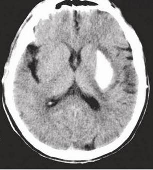
- Hypertension is the most common cause (basal ganglia, cerebellum, brainstem).
- Clinical presentation depends on region and size; may break into the ventricle.
- Intracerebral hemorrhage (ICH): hyperdense on CT; hypertensive basal ganglia hemorrhage is a master case.
Cerebral infarction (stroke)
- MCA is the most commonly affected territory.
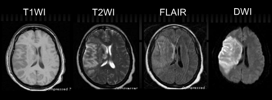
- CT: extensive low attenuation in the MCA distribution (extent); acute infarct: restricted diffusion on DWI (bright), dark on ADC.
- Old infarction → encephalomalacia with compensatory dilatation of the adjacent lateral ventricle.
- CTA/MRA to identify occlusion; perfusion for penumbra.
Aneurysm
- Abnormal focal dilation of an artery; MRA demonstrates (e.g. right MCA aneurysm near its origin); subarachnoid hemorrhage from aneurysm (basilar artery aneurysm example).
Trauma — intracranial hematomas
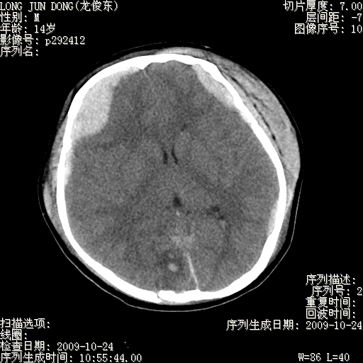
- Epidural hematoma (EDH): extra-axial blood external to the dura; lentiform (biconvex) shape; does not cross cranial sutures; associated skull fracture; often middle meningeal artery injury.
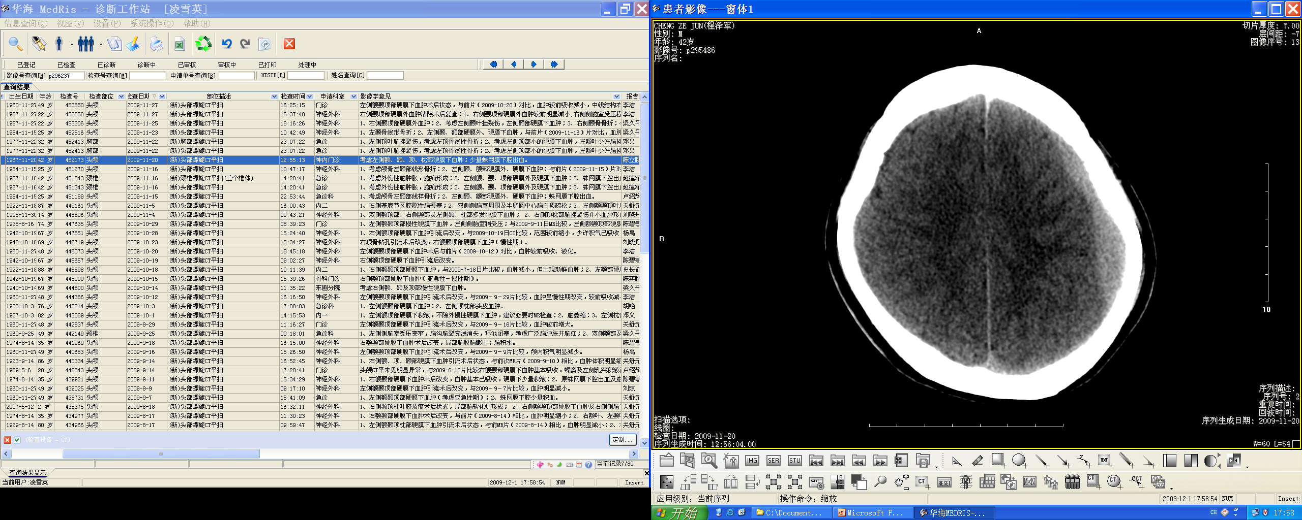
- Subdural hematoma (SDH): crescentic, beneath the dura; can cross cranial sutures (but not midline falx); acute = hyperdense crescent; chronic = hypodense; mixed density = acute + chronic components; huge SDH → midline shift + ventricular compression.
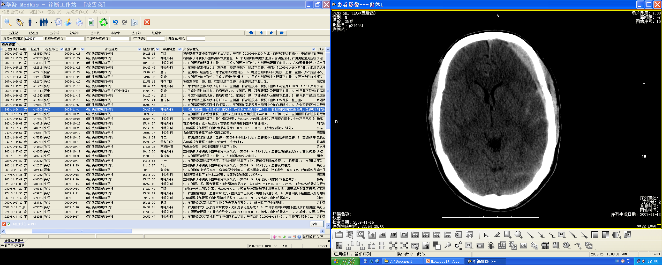
- Contusion/laceration: patchy heterogeneous high density (hemorrhage) mixed with low density (edema) — common in significant head injury.
Brain abscess
- Collection of pus enclosed within a capsule.
- MRI: well-circumscribed lobulated lesion, high signal on FLAIR; central restricted diffusion on DWI (dark on ADC); enhancing wall (ring enhancement).
- DDx from necrotic tumor: abscess = central restricted diffusion.
High-Yield Points
- Key Point: Extra-axial: meningioma (dural tail, hyperostosis) / schwannoma (CPA, IAC widening); intra-axial: metastasis / astrocytoma.
- Key Point: GBM = ring-enhancing mass crossing midline with central necrosis + edema; most common primary CNS malignancy.
- Key Point: Metastasis = gray-white junction, sphere, "small nodule large edema", 80% from lung/renal/breast/melanoma/colorectal.
- Key Point: Pituitary macroadenoma = snowman + optic chiasm compression; microadenoma = hypoenhancing on dynamic scans (hyperprolactinemia).
- Key Point: EDH = lentiform, doesn't cross sutures; SDH = crescentic, crosses sutures.
- Key Point: Acute infarct = DWI bright/ADC dark (restricted diffusion); MCA territory most common.
- Key Point: Hypertensive hemorrhage: basal ganglia/cerebellum/brainstem; hyperdense on CT.
- Key Point: Abscess = ring enhancement + central restricted diffusion.
LMCHK OSCE Practice(OSCE & LMCHK)
- CT findings of stroke and SAH are explicit LMCHK stations: ischemic stroke = low-density wedge in vascular territory (DWI confirmatory); SAH = hyperdense blood in basal cisterns/sulci from aneurysm rupture.
- EDH vs SDH is a classic discriminator: lentiform vs crescentic; crosses sutures or not.
- Meningioma: extra-axial mass + dural tail + hyperostosis → benign, WHO I.
- Metastasis vs GBM vs abscess: use DWI (abscess restricts) + location (gray-white junction vs subcortical).
- Pituitary macroadenoma compressing the optic chiasm → bitemporal hemianopia; microadenoma → hyperprolactinemia; dynamic MRI is key.
Topic Summary
Brain disease imaging: classify masses by intra- vs extra-axial location (metastasis/astrocytoma vs meningioma/schwannoma). Master cases: meningioma (dural tail + hyperostosis), GBM (ring enhancement + necrosis + midline crossing), vestibular schwannoma (CPA + IAC widening), pituitary adenoma (snowman/optic chiasm; dynamic enhancement for microadenoma), metastases (gray-white junction, small nodule large edema), hypertensive hemorrhage (basal ganglia), MCA infarction (DWI restricted), EDH (lentiform) vs SDH (crescentic), and brain abscess (ring enhancement + central restricted diffusion).
Illustrations(图解速览)
Case gallery for this chapter — identify the imaging technique, location, features and diagnosis for each: