# Ch16: MSK Imaging — Bone Tumors & Infections(骨肿瘤与感染)
Preparatory Mindset
Bone tumor imaging relies on plain film radiography as the best imaging technique for characterization; MRI or CT delineate the full extent of the tumor and its relationship to the neurovascular bundle; radionuclide bone scanning diagnoses metastatic bone disease. Decide the nature of a localized lesion using the seven features (zone of transition, adjacent cortex, expansion, periosteal reaction, calcific densities, soft tissue swelling, site), then apply the classic tumor signatures (age + site + appearance).
Core Concepts
Primary malignant bone tumors (general features)
- Plain films: poorly defined margins, destruction of the cortex, periosteal reaction, soft tissue mass; radionuclide: substantially increased activity; MRI: the most accurate technique.
Osteosarcoma
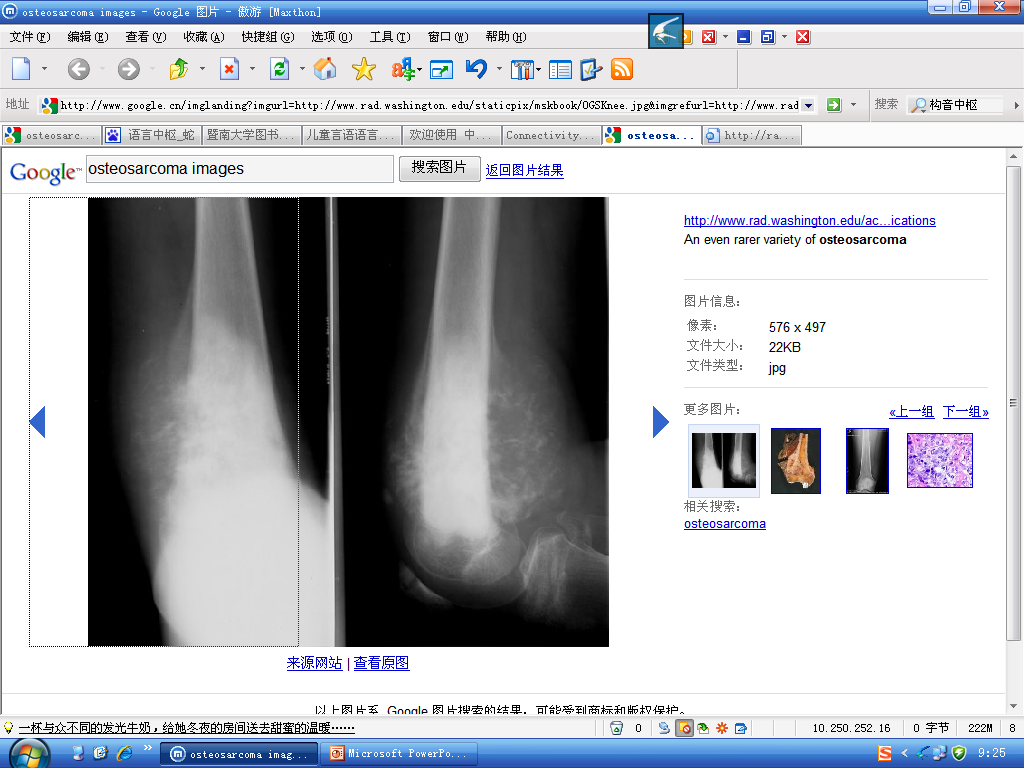
- Age: 5–20 years (or elderly following malignant change in Paget's disease).
- Site: metaphysis, most commonly around the knee (distal femur/proximal tibia).
- Imaging: bone destruction with new bone formation; florid spiculated periosteal reaction = "sunray appearance"; Codman's triangle (elevated periosteum at tumor edge).
Chondrosarcoma
- Age: 30–50 years.
- Site: pelvis, scapulae, humeri, femora.
- Imaging: lytic expanding lesion; flecks of calcium (cartilage origin); large extraosseous soft tissue component best on CT/MRI; periosteal reaction usually present; may arise from malignant degeneration of a benign cartilage tumor.
Ewing's sarcoma
- Highly malignant; commonest in children.
- Site: shaft (diaphysis) of long bones.
- Imaging: ill-defined bone destruction; "onion skin" periosteal reaction.
Giant cell tumor
- Has features of both malignant and benign tumors.
- Site: around the knee or wrist AFTER epiphyses have fused (young adults, 20–40).
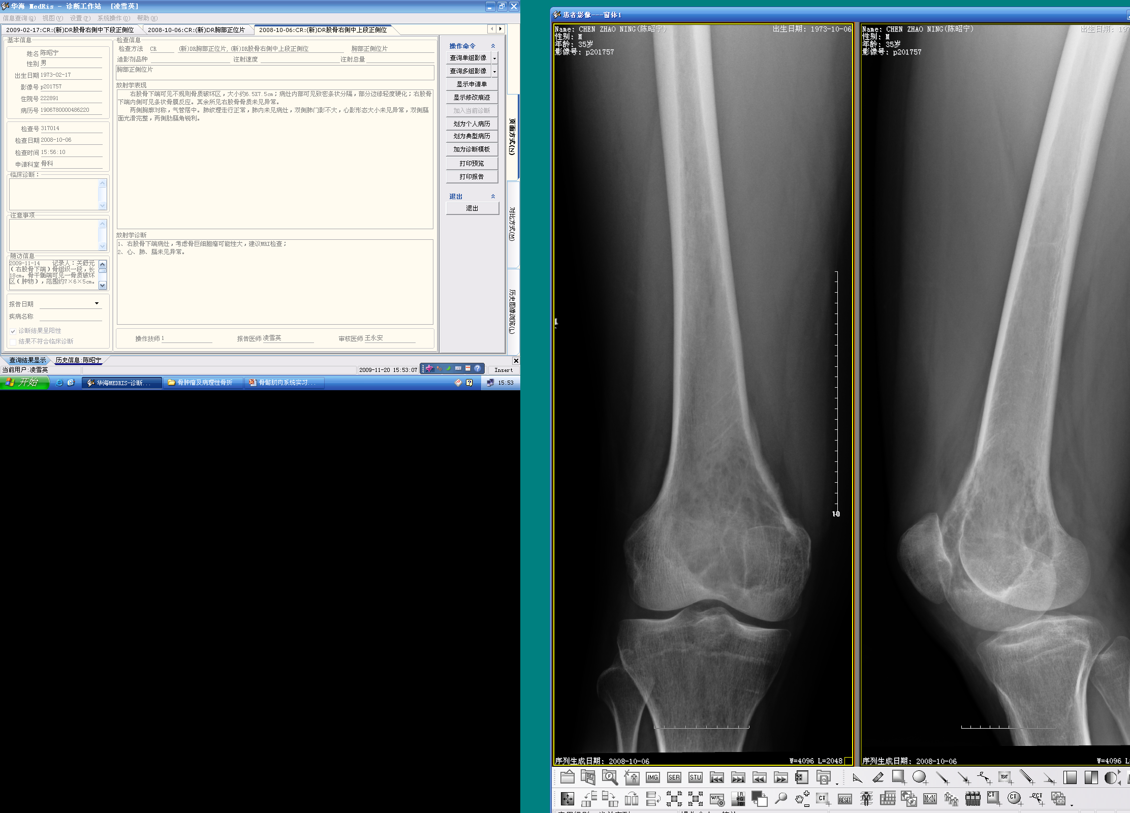
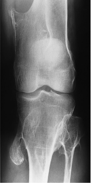
- Imaging: eccentric expanding destructive lesion, subarticular in position; fairly well defined margin; thin cortex, may be completely destroyed; strands of bone crossing the lesion.
Benign tumors and tumor-like conditions
- General: well-demarcated edge with a sclerotic rim; cause expansion but rarely breach the cortex; no soft tissue mass; periosteal reaction unusual unless fracture; radionuclide little/no increased activity.
- Enchondroma: lytic expanding lesion (often in small bones of hands/feet); popcorn calcification.
- Fibrous dysplasia: expansion with intact cortex; ground-glass density.
- Osteoid osteoma: small lytic nidus with surrounding sclerosis; night pain relieved by aspirin; uncertain nature.
- Osteochondroma: cartilage-capped bony outgrowth (exostosis); metaphyseal, away from joint.
- Eosinophil granuloma (Langerhans' histiocytosis): children/young adults; lytic lesions in skull ("geographic skull"), pelvis, femur, ribs; may look aggressive or well-defined with sclerotic rim.
- Bone cyst (simple): well-defined lytic lesion in the proximal humerus/femur in children.
Osteomyelitis
- Often Staphylococcus aureus; affects infants and children.
- Early radiographs normal — bone changes not visible until 10–14 days after onset; radionuclide bone scan and MRI show changes within a day or two.
- Affects the metaphysis of a long bone (femur/tibia); earliest signs: soft tissue swelling, bone destruction; periosteal reaction may become extensive forming an involucrum; sequestra (dead bone) and sinus tracks.
- Chronic stage: bone thickened and sclerotic, loss of cortex-medulla differentiation; Brodie's abscess (lucency in tibia surrounded by sclerosis).
- MRI is the investigation of choice (bone edema + pus); US demonstrates subperiosteal pus before plain-film changes.
Metastases (the commonest malignant bone tumor)
- May be sclerotic, lytic, or mixed.
- Bones containing red marrow most frequently affected: spine, skull, ribs, pelvis, humeri, femora.
- Lytic metastases in adults: breast, prostate, bronchus; in children: neuroblastoma, leukemia. Lytic = well/ill-defined destruction without sclerotic rim.
- Metastases and myeloma are virtually the only causes of multiple obvious lytic lesions in bone.
- Sclerotic metastases: ill-defined areas of increased density (prostate, breast, lung).
- Radionuclide bone scan: much more sensitive than plain films (numerous areas of increased uptake with known primary = virtually certain diagnosis; low specificity). MRI better than radionuclide scan (shows more lesions; whole-spine MRI for metastatic survey/myeloma; DWI for detection; important when bone scan normal/equivocal with strong suspicion, or suspected cord compression). CT less sensitive than MRI.
Multiple myeloma
- Most frequently in bones with active hemopoiesis; lesions may resemble lytic metastases but are often better defined and may cause expansion; diffuse marrow involvement → generalized loss of density (mimics osteoporosis); full skeletal survey with plain radiography is a standard staging tool.
Generalized bone density changes
- Osteoporosis / osteomalacia / hyperparathyroidism / myeloma → generalized decrease.
- Osteopetrosis (marble bone), myelosclerosis, sclerotic metastases → generalized increase.
Paget's disease
- Chance finding in the elderly; pelvis, spine, skull, long bones; bone softening causes bowing, deformity, pathological fractures; coarse trabeculae + thickened cortex + enlarged bone; malignant degeneration (osteosarcoma) is an occasional complication.
Hemolytic anemia (marrow hyperplasia)
- Thalassaemia, sickle cell: marrow hyperplasia → thinned cortex, resorbed trabeculae, generalized decreased density; skull: widened diploë + "hair-on-end" striations; ribs enlarged; phalanges rectangular; sickle cell: infarction and infection.
Joint diseases (lab)
- Rheumatoid arthritis: erosions, periarticular osteoporosis, joint space narrowing (hands/wrists).
- Osteoarthritis: joint space narrowing, osteophytes, subchondral sclerosis.
- Avascular necrosis: femoral head flattening/sclerosis (MRI best).
- Gout: punched-out erosions with overhanging edges; septic/pyogenic and tuberculous arthritis: joint space loss + bone destruction.
High-Yield Points
- Key Point: Plain film = best for bone tumor characterization; MRI for extent; bone scan for metastases.
- Key Point: Osteosarcoma = metaphysis around knee, 5–20 y, sunray + Codman's triangle.
- Key Point: Ewing's = diaphysis, children, onion-skin periosteal reaction.
- Key Point: Chondrosarcoma = pelvis, 30–50 y, flecks of calcium.
- Key Point: Giant cell tumor = subarticular, post-epiphyseal-fusion, eccentric expansion (knee/wrist).
- Key Point: Osteomyelitis = metaphysis, S. aureus, plain films negative first 10–14 days, MRI/bone scan early.
- Key Point: Metastases commonest malignant bone tumor; spine/skull/ribs/pelvis/femur/humeri; lytic/sclerotic/mixed.
- Key Point: Metastases + myeloma = only causes of multiple obvious lytic lesions.
- Key Point: Bone scan sensitive but nonspecific; MRI best overall (whole-spine survey, cord compression).
- Key Point: Paget's = coarse trabeculae + bone enlargement; rare osteosarcoma degeneration.
LMCHK OSCE Practice(OSCE & LMCHK)
- Femoral shaft tumour (tumour x-ray) is a listed LMCHK station: describe location (diaphysis), zone of transition, cortical destruction, periosteal reaction (sunray/onion skin), soft tissue mass → give differential (osteosarcoma vs Ewing's vs metastasis) based on age.
- Osteosarcoma vs Ewing's: age (5–20 vs child) + site (metaphysis vs diaphysis) + periosteal reaction (sunray vs onion-skin) — classic discriminator.
- Bone metastasis survey: patient with known primary (breast/prostate/lung) + bone pain → bone scan (hot spots) or MRI; sclerotic = prostate/breast; lytic = lung/thyroid/renal; "multiple lytic lesions = metastasis or myeloma."
- Septic arthritis/osteomyelitis in a child: limp + fever → plain films may be normal early; MRI/bone scan confirm; S. aureus.
Topic Summary
Bone tumors: plain film for characterization (7 features), MRI/CT for extent, bone scan for mets. Malignant signatures: osteosarcoma (metaphysis, sunray, Codman's, 5–20 y), chondrosarcoma (pelvis, flecks of calcium, 30–50 y), Ewing's (diaphysis, onion-skin, child), giant cell tumor (subarticular, eccentric). Osteomyelitis: metaphysis, S. aureus, delayed plain-film changes, MRI/bone scan early. Metastases (commonest): red-marrow bones, lytic/sclerotic/mixed, bone-scan sensitive; with myeloma = the two causes of multiple lytic lesions. Metabolic/generalized: osteoporosis, osteomalacia, Paget's, marrow hyperplasia.
Illustrations(图解速览)
Case gallery for this chapter — identify the imaging technique, location, features and diagnosis for each: