# Ch09: Hepatobiliary & Pancreatic Imaging(肝胆胰影像)
Preparatory Mindset
Imaging of the liver, biliary tree and pancreas uses plain film (acute abdomen), ultrasound (screening), CT and MRI with multiphasic contrast enhancement. The key teaching points: normal anatomy (liver 8 segments; pancreatic duct diameters), diffuse liver disease (fatty liver, cirrhosis), focal liver lesions (cyst, hemangioma, HCC, metastasis) with their enhancement patterns, biliary disease (dilatation, choledocholithiasis, cholangiocarcinoma), and pancreatic disease (acute/chronic pancreatitis, pancreatic carcinoma). The area of difficulty: interpreting enhancement patterns on multi-phase CT/MRI.
Core Concepts
Imaging techniques
- Plain film: acute abdomen assessment.
- US: screening; first-line for gallstones; pros: no radiation, real-time, Doppler; cons: operator-dependent, limited by bowel gas.
- CT: multiphasic contrast-enhanced scans are crucial — non-contrast, arterial (~25–30 s), portal venous (~60–70 s), delayed/equilibrium (2–5 min); pros: fast, comprehensive, calcification/hemorrhage/vascular mapping/staging; cons: radiation, contrast allergy.
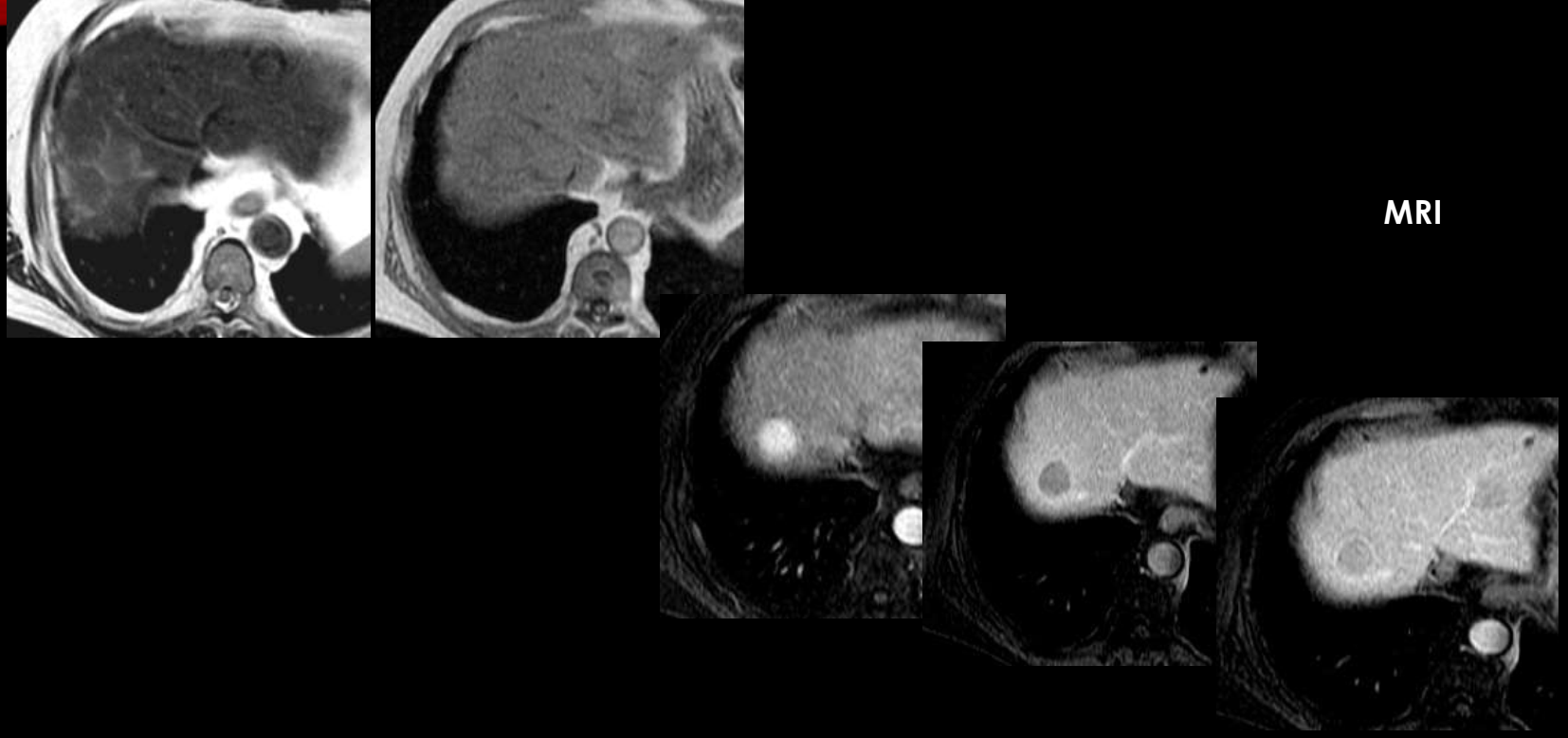
- MRI: T1W, T2W, DWI, MRCP, multiphasic contrast; ideal for focal liver lesion characterization; MRCP non-invasively visualizes the biliary tree and pancreatic duct.
- Liver receives dual blood supply: 25% hepatic artery, 75% portal vein.
Normal anatomy
- Liver divided into eight segments (Couinaud), each with independent vascular supply and biliary drainage. Porta hepatis = entrance/exit of major vessels and bile ducts.
- Key vessels: hepatic veins (RHV/MHV/LHV → IVC), portal vein (main trunk, right/left branches), splenic vein, SMV/SMA, celiac axis.
- Pancreas: head, body, tail; pancreatic duct normal 1–3.5 mm (head 3.5 mm, body 2.5 mm).
- Biliary dilatation thresholds: intrahepatic ducts >40% of adjacent portal vein diameter; common duct >8 mm; gallbladder >5 cm.
Diffuse liver disease
- Fatty liver (steatosis): accumulation of fat in liver cells. Normal adult: liver attenuation consistently higher than spleen. Mild diffuse steatosis: liver < spleen attenuation. Marked: liver lower than hepatic blood vessels. MRI in/out-of-phase: signal drop on out-of-phase; focal fat has no mass effect (vessels run normal course).
- Cirrhosis: extensive scarring from chronic injury. Imaging: hypertrophy of caudate and left lobes with shrinkage of the right lobe; inhomogeneous parenchyma; nodular liver surface; extrahepatic signs of portal hypertension (splenomegaly, ascites, varices, portosystemic collaterals, gallbladder wall thickening, Gamna-Gandy bodies in spleen).
Focal liver lesions
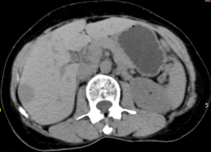
- Simple cyst: well-circumscribed, homogeneous, near-water attenuation (<20 HU); no enhancement; hypointense T1, hyperintense T2; no discernible wall.
- Cavernous hemangioma (most common benign liver tumor): fed by hepatic artery branches, slow internal circulation, stable size. CT: well-defined hypodense on unenhanced; nodular peripheral enhancement progressing toward the center (globular, peripheral); equilibrium phase: near-complete fill-in isodense to IVC blood; T2WI: marked hyperintensity ("light bulb" sign). Do not need contrast if typical on USG.
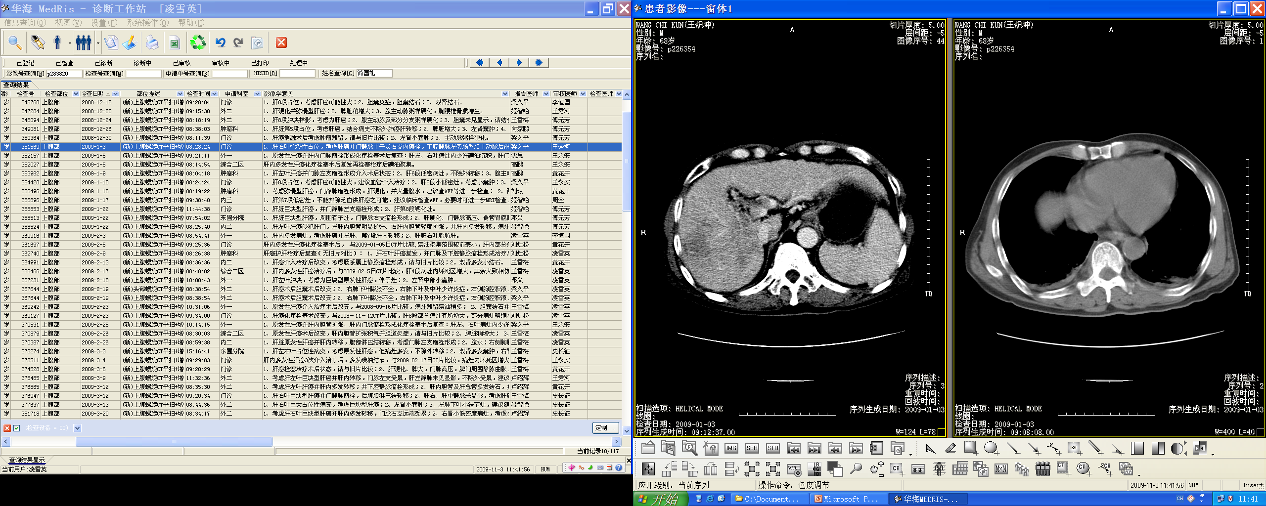
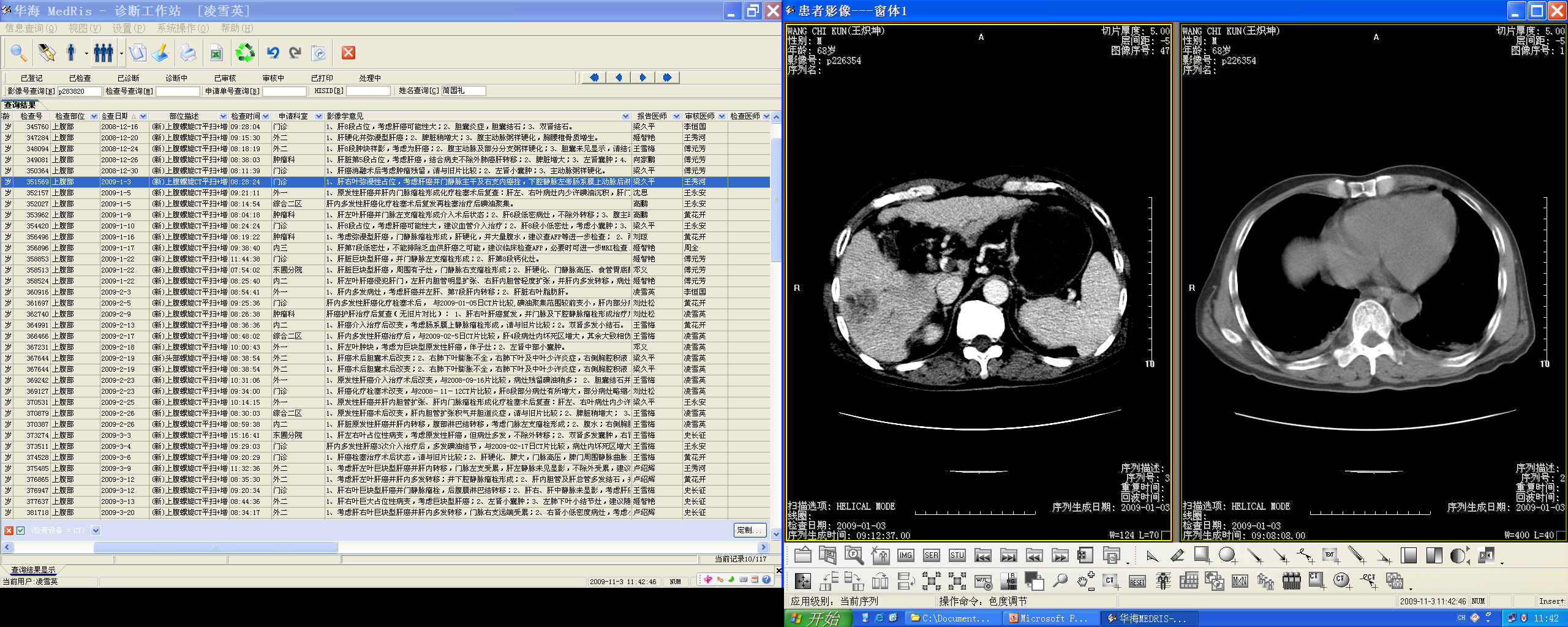
- Hepatocellular carcinoma (HCC) (most common primary malignancy): risk factors cirrhosis, chronic hepatitis; growth patterns: solitary massive, multinodular, diffuse infiltrative; AFP often elevated. Hypervascular on arterial phase with washout on portal venous phase; tumor capsule (sharply marginated rim); tumor thrombus (portal vein filling defect); central necrosis; satellite lesions.
- Metastases (most common malignant masses in liver): originate from GI tract, breast, lung; multiple; hypoattenuating with continuous ring-like (rim) enhancement; "bull's eye" sign; necrosis/fibrosis/calcification/hemorrhage.
- HKHA/HK reference (LI-RADS-based approach): chronic liver disease — subcentimetric lesion (<1 cm) → repeat USG in 3 months; lesion ≥1 cm → contrast cross-sectional imaging (triphasic/4-phase CT, MRI with gadolinium or gadoxetate (Primovist), or dual-tracer PET-CT in selected patients); liver biopsy only if imaging non-diagnostic (MDT decision).
Biliary disease
- Choledocholithiasis: ~20% of adult obstructive jaundice; CT high-density calcification within the duct; MRCP sensitivity 86–100%, specificity 85–100% for ductal stones.
- Cholangiocarcinoma (bile duct epithelial adenocarcinoma): growth patterns — mass forming (delayed enhancement, biliary dilatation, atrophy), periductal infiltrating (focal circumferential duct thickening + proximal dilatation), intraductal polypoid.
Pancreatic disease
- Pancreatic carcinoma (highly lethal): CT recommended for initial assessment; hypodense mass distorting the gland contour; obstruction of CBD + pancreatic duct + distal atrophy; double duct sign (dilated CBD + pancreatic duct); signs of unresectability: adjacent organ involvement, regional nodes >15 mm, peripancreatic vessel encasement/obstruction, liver metastases, peritoneal carcinomatosis.
- Acute pancreatitis: alcohol or obstructing gallstone; two types — interstitial edematous (90–95%) and necrotizing. Findings: focal/diffuse parenchymal enlargement, density change from edema, indistinct margins, retroperitoneal fat stranding; necrosis = lack of parenchymal enhancement (best evaluated 48–72 h after onset). Complications: pancreatic necrosis, fluid collections, pseudocyst (fibrous wall, no epithelial lining; internal dependent debris on MR is specific), pancreatic abscess (thicker irregular walls, gas locules).
- Chronic pancreatitis (most commonly alcohol): irreversible damage; calcifications in the distribution of the pancreatic duct are pathognomonic; coarse/punctate calcifications across the LUQ.
High-Yield Points
- Key Point: Multiphasic CT: arterial 25–30 s, portal venous 60–70 s, delayed 2–5 min.
- Key Point: HCC = arterial hyperenhancement + portal venous washout + capsule + tumor thrombus.
- Key Point: Hemangioma = peripheral nodular enhancement with delayed fill-in; "light bulb" bright on T2.
- Key Point: Metastases = multiple, rim enhancement, bull's-eye.
- Key Point: Cyst = water density <20 HU, no enhancement, no wall.
- Key Point: Cirrhosis = right lobe shrinkage + caudate/left hypertrophy + nodular surface + portal HTN stigmata.
- Key Point: Double duct sign = pancreatic head carcinoma.
- Key Point: Acute pancreatitis: interstitial edematous vs necrotizing; necrosis = non-enhancement; pseudocyst vs abscess.
- Key Point: Chronic pancreatitis = pancreatic duct distribution calcification (pathognomonic).
- Key Point: ≥1 cm liver lesion in chronic liver disease → contrast cross-sectional imaging (LI-RADS approach).
LMCHK OSCE Practice(OSCE & LMCHK)
- HCC is an LMCHK common case (HBP): describe the multiphasic enhancement (arterial enhancement, washout), cirrhosis background, portal vein thrombus; imaging choice: triphasic CT or MRI; AFP; biopsy only if non-diagnostic (MDT).
- "CT abdomen of adrenal glands" is a listed ECS station; adrenal mass characterization uses washout CT.
- Jaundice workup: US first (biliary dilatation) → MRCP for stones/strictures → contrast CT/MRI for mass; double duct sign → pancreatic head tumor.
- Pancreatic carcinoma: CT hypodense mass + double duct sign + unresectability features (vessel encasement, liver mets).
Topic Summary
Hepatobiliary-pancreatic imaging relies on multiphasic contrast CT/MRI. Diffuse disease: fatty liver (attenuation < spleen; out-of-phase drop) and cirrhosis (right-lobe atrophy, caudate hypertrophy, nodular surface, portal HTN). Focal lesions are separated by enhancement: cyst (none), hemangioma (peripheral nodular → fill-in, bright T2), HCC (arterial ↑ → venous washout, capsule, thrombus), metastases (rim enhancement). Biliary: choledocholithiasis (MRCP), cholangiocarcinoma (delayed enhancement/infiltrating). Pancreas: carcinoma (hypodense + double duct), acute pancreatitis (interstitial vs necrotizing; pseudocyst vs abscess), chronic pancreatitis (ductal calcification).
Illustrations(图解速览)
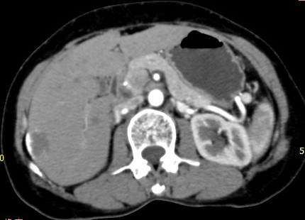
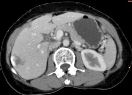
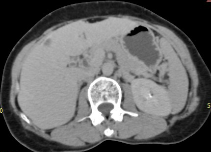
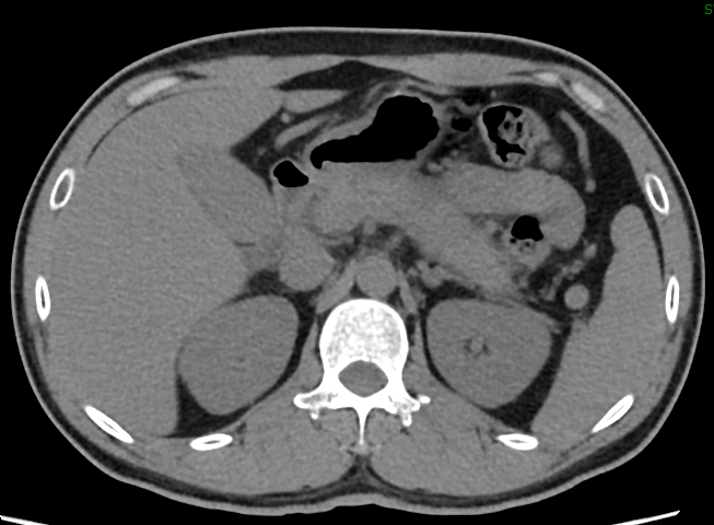
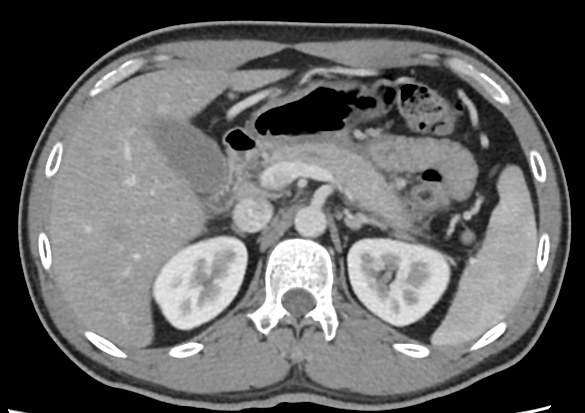
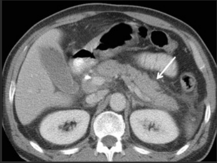
Case gallery for this chapter — identify the imaging technique, location, features and diagnosis for each: