# Ch06: Cardiovascular Imaging(心血管影像)
Preparatory Mindset
Cardiac imaging provides information about: structure and mechanical function of chambers and valves, myocardial tissue characteristics (scar/edema), stress-induced reversible or irreversible perfusion defects, and coronary artery patency. Plain chest X-ray is mandatory as the first investigation in cardiac disease — it assesses the effects of cardiac disease on the lungs and pleural cavities and gives limited information about the heart itself. Echocardiography is the first-line modality for exploring heart disease; CT/CTA, MRI/MRA and DSA serve specific roles.
Core Concepts
Modalities
- CXR: assess size and shape, outline of heart and great vessels, valvular/pericardial calcifications, and — critically — the pulmonary vasculature. Views: PA, lateral, RAO, LAO.
- Echocardiography (US): first-line; morphological and functional info (valve anatomy/motion, chamber volume, wall thickness, blood flow velocity/direction); first-line for pericardial disease/effusion.
- CT & CTA: heart valves, muscles, great vessels; chamber morphology/volume; wall thickness; cardiac/pericardial calcification; thrombus; pulmonary embolism; coronary arteries; dissection.
- MRI & MRA: complex congenital heart disease, myocardial thickness/disease, great vessels (dissecting aneurysm), pericardial disease; MRA without contrast.
- DSA (cardiac catheterization): catheters into heart/vessels with contrast (coronary angiography); Seldinger method.
Heart borders on CXR
- PA view borders: ① superior vena cava; ② right atrium; ③ aortic arch; ④ main pulmonary artery; ⑤ left atrial appendage; ⑥ left ventricle.
- Lateral view borders: ① trachea; ② right ventricle; ③ left ventricle; ④ left atrium; ⑤ right pulmonary artery.
- 5 important cardiac contours: ascending aorta, aortic knob, main pulmonary artery segment, the indentation where left atrial enlargement appears on the left, and the double density of left atrial enlargement on the right.
Normal variants affecting heart shape
- Body build: transverse / oblique / long narrow heart.
- Age: infant → globular; adult → oblique/horizontal with age.
- Respiration: inspiration drops and narrows the heart; expiration makes it horizontal.
- Position: erect vs supine.
Heart size — cardiothoracic ratio (CTR)
- CTR = (transverse diameter of heart) ÷ (maximum internal transverse diameter of chest); normal ≤ 0.5 in adults.
- Measure maximal diameter from midline to left border (b) + diameter to right border (a); should not exceed half the maximum thoracic diameter (c).
Chamber enlargement
- Left ventricular enlargement: cardiac apex extends left and down; retrocardiac space narrowed/disappears; LV segment extended, rounded, expanded left; aorta prominent. Causes: hypertension, aortic valve disease, mitral incompetence, VSD, PDA.
- Left atrial enlargement: double density on the right border (PA); indentation on the left border (left atrial appendage dilatation); esophagus displaced posteriorly (lat/RAO); elevation of left mainstem bronchus (LAO); widening of carinal angle. Causes: mitral lesion, LV failure, PDA, VSD.
- Right atrial enlargement: enlargement/bulging of the right heart border (convex border projecting abnormally far into the right lower lung field; RA exceeds lower half of right mediastinal border). Causes: right ventricular failure, tricuspid disease.
- Right ventricular enlargement: MPA prominent; lateral: contact between front of heart and sternum >1/3 (narrowed retrosternal space); elevated/rounded apex. Causes: mitral stenosis, chronic pulmonary heart disease, pulmonary stenosis, pulmonary HTN, Fallot's tetralogy, ASD, VSD.
Pulmonary vasculature — five states
| State | RDPA (right descending PA) | Upper vs lower lobe vessels | Pattern |
|---|---|---|---|
| Normal | ≤15–17 mm | Lower > upper | Gradual tapering central→peripheral |
| Pulmonary venous HTN | usually >17 mm | Upper = or ≥ lower (cephalization); upper zone vessels >3 mm = earliest change | — |
| Pulmonary arterial HTN | — | — | Pruning (rapid cutoff of peripheral vessels; central vessels too large) |
| Increased flow (plethora) | usually >15 mm | All vessels everywhere bigger (distribution maintained, lower > upper) | — |
| Decreased flow (oligemia) | small | Fewer vessels, small hila | — |
- Plethora = increased pulmonary blood flow (enlargement of pulmonary vessels); oligemia = decreased flow (general reduction in width of pulmonary arteries).
Pericardial disease
- Pericardial effusion: heart shadow greatly enlarged with no specific chamber enlargement; marked change in transverse diameter within 1–2 weeks without pulmonary edema; globular/flask-like heart; shortening of the aortic shadow. Echocardiography first-line.
- Constrictive pericarditis (complication of pericarditis, particularly TB): straight borders, triangular/polygonal shape; indistinct outline (mediastinal-pleural involvement); pericardial calcification (best on lateral view, maximal along anterior and inferior borders — makes diagnosis certain); lungs normal or oligemic.
Aortic disease
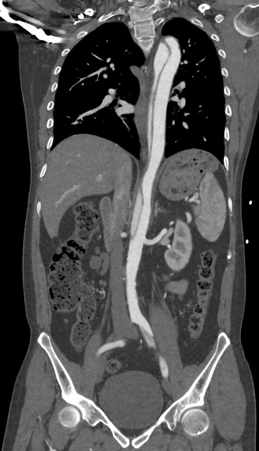
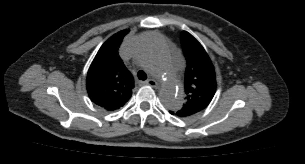
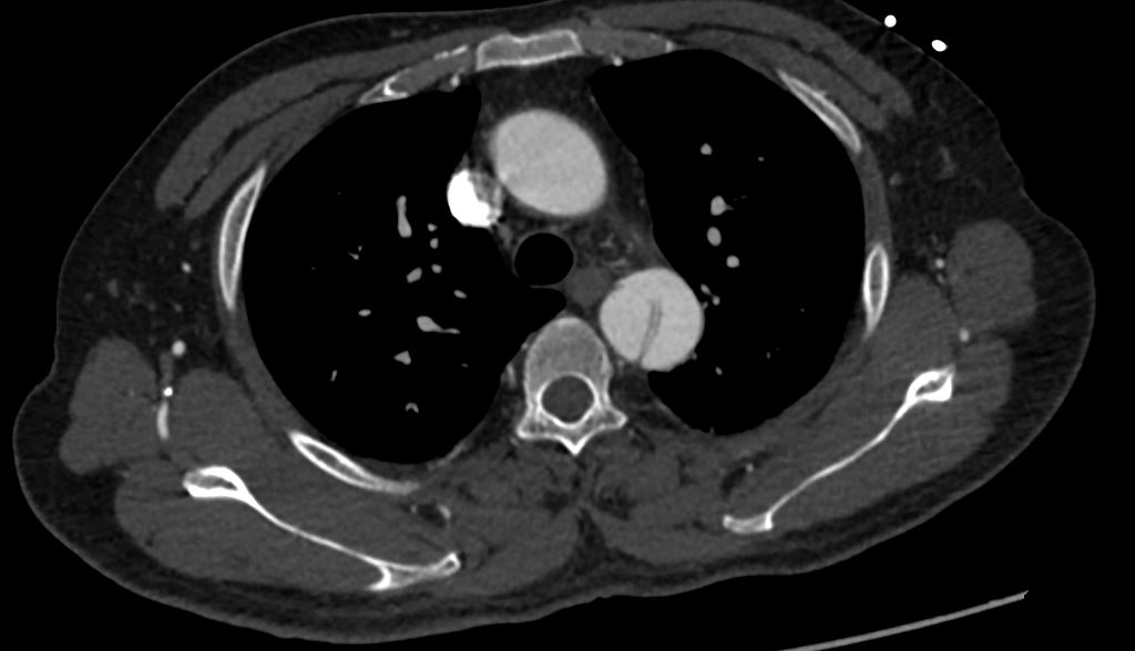
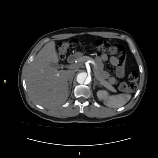
- Stanford: Type A = ascending aorta involved (regardless of primary tear); Type B = descending only. - DeBakey: Type I = ascending + descending; Type II = ascending only; Type III = descending only (mnemonic BAD: B = both, A = ascending, D = descending). - Diagnosis: NCCT shows displaced calcified intimal flap; CTA shows intimal tear, true vs false lumen.
- Aorta shape/density changes: elongation, widening, calcification; aortic knob high (above clavicle sometimes); demarcation between ascending aorta and right atrium descends.
- Dissection of aorta (medical emergency): sudden severe tearing chest-back pain radiating to neck/abdomen; hemorrhage in the media separates media from adventitia forming a pseudocoele.
- Aneurysm thresholds: ascending aorta >5 cm; descending aorta >4 cm; abdominal aorta >3 cm (normal abdominal aorta after 50 y ≈ 2 cm). >6 cm ascending / >5 cm abdominal = high risk of rupture. Ruptured AAA triad: pain, hypotension, pulsatile abdominal mass.
Specific cardiac disorders
- Heart failure: cardiac enlargement + pulmonary venous hypertension + pulmonary edema + pleural effusion.
- Mitral stenosis: LA → PV → PA → RV (mitral valve type heart); pulmonary venous HTN with left atrial + right ventricular enlargement; small LV and aortic knuckle; Kerley B lines (interstitial edema); MPA prominent.
- Congenital heart disease: ASD, VSD, PDA (increased flow/plethora); PS; Tetralogy of Fallot (oligemia).
Coronary artery disease
- Coronary atherosclerosis: stenosis grading 0% / <50% mild / 50–75% moderate / >75% severe / 100% blocked; CTA of coronary arteries; coronary calcification on CT.
High-Yield Points
- Key Point: CXR is the mandatory first investigation in cardiac disease; echo is first-line for exploring heart disease.
- Key Point: CTR ≤ 0.5 (transverse heart diameter ÷ internal thoracic diameter).
- Key Point: LA enlargement = double density (right) + indentation (left) + esophageal displacement + left main bronchus elevation.
- Key Point: Pulmonary venous HTN = cephalization (upper ≥ lower lobe vessels, upper zone >3 mm); PAH = pruning; plethora = everything big; oligemia = small hila.
- Key Point: Aortic dissection: Stanford A (ascending) vs B (descending); DeBakey I/II/III (BAD mnemonic); CTA is diagnostic.
- Key Point: Aneurysm: ascending >5 cm, descending >4 cm, abdominal >3 cm; >6 cm (asc) / >5 cm (abd) high rupture risk.
- Key Point: Constrictive pericarditis = pericardial calcification (lateral view) + abnormal triangular cardiac shape.
LMCHK OSCE Practice(OSCE & LMCHK)
- Aortic dissection (DeBakey III/Stanford B) is a standard case: 59 M tearing back pain → CTA shows intimal flap with true/false lumen; describe displaced calcified intima on NCCT first.
- Pulmonary embolism: CT pulmonary angiography (CTPA) is the diagnostic test; CXR may be normal or show oligemia.
- Abdominal aortic aneurysm rupture triad (pain, hypotension, pulsatile mass) → emergency; CT/CTA confirms.
- Coronary CTA stenosis grading (0/<50%/50–75%/>75%/100%) may be asked in the imaging station.
Topic Summary
Cardiovascular imaging starts with CXR (heart borders, CTR, chamber enlargement, pulmonary vasculature states) and echocardiography, with CT/CTA for coronary arteries, PE, dissection and aneurysm, MRI/MRA for congenital and myocardial disease, and DSA for coronary angiography. Master the five pulmonary vasculature states (normal, venous HTN/cephalization, arterial HTN/pruning, increased flow, decreased flow), the four-chamber enlargement patterns, pericardial disease signs (effusion = globular heart; constrictive = calcification), aortic dissection classification (Stanford/DeBakey) and aneurysm size thresholds.
Illustrations(图解速览)
Case gallery for this chapter — identify the imaging technique, location, features and diagnosis for each: