# Ch08: Acute Abdomen Imaging(急腹症影像)
Preparatory Mindset
The acute abdomen = a variety of illnesses causing acute abdominal pain from ischemia, infection or inflammation. The imaging approach is algorithmic: Is there pneumoperitoneum? → think perforation. Is there bowel dilation / a transition point? → think obstruction (find the cause). Are there signs of ischemia (pneumatosis, portal venous gas, non-enhancing bowel)? → this is a SURGICAL EMERGENCY. Modalities: abdominal X-ray (rapid screening), ultrasound (children/pregnancy/RUQ/gynecology), CT (gold standard for most non-obstetric acute abdomens).
Core Concepts
Imaging modalities
- Plain radiography (X-ray): rapid screening for pneumoperitoneum, obstruction, foreign bodies. Views: erect CXR (free air) + supine/erect AXR. Limited sensitivity.
- The ABCs of reading an AXR: A — Air (bowel gas pattern, free air, abnormal air collections); B — Bony (spine, ribs, pelvis fractures); C — Calcifications (renal/ureteric/gallstones, calcified structures).
- Ultrasound: first-line in children, pregnant women, right upper quadrant/gynecological pain; excellent for appendicitis (children), cholecystitis, AAA. No radiation, real-time.
- CT: gold standard for most non-obstetric acute abdomens; highest sensitivity/specificity; IV contrast essential; oral/rectal contrast situation-dependent; visualizes entire abdomen + retroperitoneum + abdominal wall.
Normal AXR landmarks
Hepatic angle, splenic angle, renal shadows, psoas muscle, properitoneal fat strip; normal bowel gas: stomach/duodenum gas, colon (mottled stool pattern), few small-bowel gas collections.
Calcifications on AXR
Gallstones (15–20% contain enough calcium to be visible; RUQ; laminated appearance), porcelain gallbladder (wall calcification → chronic obstruction + increased gallbladder carcinoma risk), urinary calculi (85% visible on plain film; staghorn calculus fills the collecting system), calcified adrenal glands (newborn hemorrhage, TB, Addison), pancreatic calcifications (chronic alcohol-induced pancreatitis; coarse punctate; extend across LUQ).
Bowel dilation thresholds
- Small bowel dilated > 2.5–3.0 cm; colon > 5 cm; cecum > 8 cm.
Mechanical small bowel obstruction (SBO)
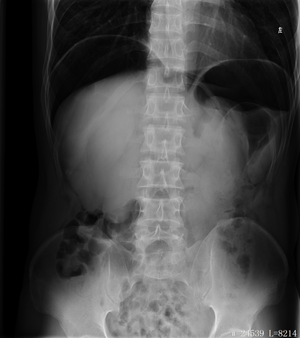
- Dilated loops of small bowel (>3 cm); air-fluid levels >2.5 cm long; air-fluid levels at differing heights within the same loop (stair-step sign — strong evidence of obstruction); small bubbles of gas trapped between valvulae conniventes; colonic collapse; paucity of air in colon/rectum.
- Causes: adhesions (most common), hernias, tumors, intussusception, Crohn stricture, gallstone ileus, volvulus.
- Erect film: air-fluid levels at different heights; valvulae conniventes extend across the entire lumen diameter.
Mechanical large bowel obstruction (LBO)
- Most colonic obstructions in the sigmoid colon.
- Dilation of colon from cecum to the point of obstruction; paucity/absence of gas distal; air-fluid levels in dilated colon (upright); small bowel distention if ileocecal valve incompetent.
- Causes: carcinoma (commonest), diverticulitis, volvulus, stricture.
Sigmoid volvulus
- Large gas-filled loop (inverted U / coffee-bean shape) without haustra, arising from the pelvis and extending high into the abdomen, often to the diaphragm; no gas in the rectum; barium enema shows beaking sign at the twist; CT: "whirl" of the mesentery at the point of volvulus.
Adynamic (paralytic) ileus
- Reduced peristalsis without mechanical obstruction; decreased/absent peristalsis; less intraluminal fluid; diffuse gaseous distension of small bowel and colon/rectum (gas in the rectum favors ileus over obstruction).
Critical imaging signs
- Pneumoperitoneum (free air): indicates hollow viscus perforation. X-ray: free air under the diaphragm (erect view); CT: free air outlining the abdominal wall or between bowel loops. Common causes: GI perforation, trauma, recent surgery.
- Pneumatosis intestinalis & portal venous gas: critical sign of bowel ischemia/infarction — gas within the bowel wall or portal venous system.
Bowel ischemia (surgical emergency)
Signs: bowel wall thickening/edema; hypoenhancement or non-enhancement; mesenteric fat stranding/fluid; vascular thrombosis (SMV/SMA); pneumatosis intestinalis & portal venous gas; strangulated SBO (adhesions/volvulus); decreased enhanced small bowel in ischemia.
Perforation & acute abdomen summary
- Pneumoperitoneum → PERFORATION.
- Bowel dilation/transition point → OBSTRUCTION (find the cause).
- Pneumatosis, portal gas, non-enhancing bowel → SURGICAL EMERGENCY.
High-Yield Points
- Key Point: Erect CXR is the best plain-film test for free air (under the diaphragm).
- Key Point: SBO: dilated small bowel >3 cm, stair-step air-fluid levels, collapsed colon/rectum.
- Key Point: LBO: dilated colon to the transition point, absent gas distally; sigmoid commonest site.
- Key Point: Sigmoid volvulus = coffee-bean/inverted-U gas loop + no rectal gas + beaking on enema.
- Key Point: Paralytic ileus = gas throughout including rectum, no transition point.
- Key Point: Pneumatosis + portal venous gas = ischemia/infarction = surgical emergency.
- Key Point: CT with IV contrast = gold standard for acute abdomen; US first-line in children/pregnancy.
LMCHK OSCE Practice(OSCE & LMCHK)
- "Crescent-shaped low density under the right hemidiaphragm on an erect chest film" → pneumoperitoneum (GI perforation) — a classic station; state the diagnosis and the need for urgent surgery.
- Small bowel obstruction: describe dilated small bowel loops with valvulae conniventes + air-fluid levels at different heights; give causes (adhesions, hernia, tumor).
- Sigmoid volvulus: recognize the coffee-bean sign and inverted-U configuration; no gas in the rectum.
- Always mention the best next imaging test: CT with IV contrast for non-obstetric acute abdomen; US for children/pregnant women.
Topic Summary
The acute abdomen is approached with a three-question algorithm: perforation (pneumoperitoneum on erect CXR/CT), obstruction (dilated loops + transition point + air-fluid levels; SBO vs LBO vs volvulus vs ileus), and ischemia (pneumatosis, portal venous gas, non-enhancing bowel — surgical emergency). AXR uses the ABCs (air, bony, calcifications); US is first-line for children/pregnancy; CT with IV contrast is the gold standard for most non-obstetric acute abdomens.
Illustrations(图解速览)
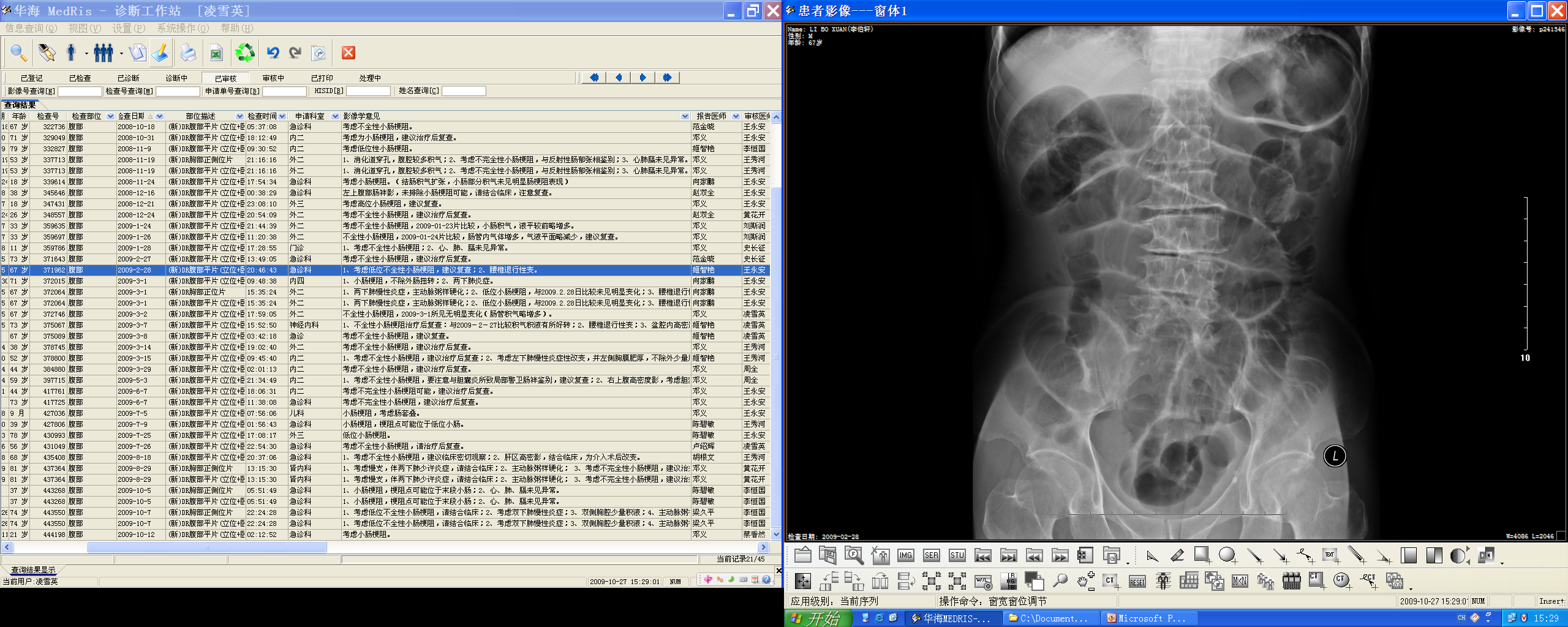
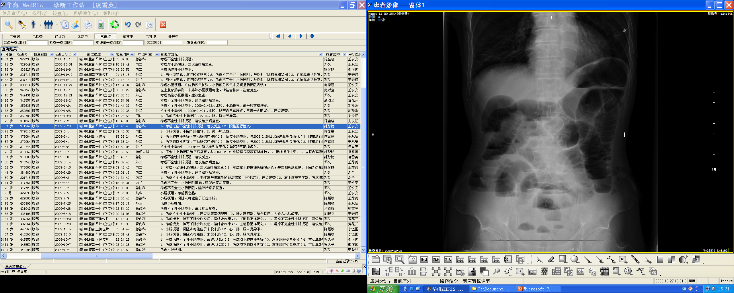
Case gallery for this chapter — identify the imaging technique, location, features and diagnosis for each: