# Ch10: Genitourinary Imaging(泌尿生殖影像)
Preparatory Mindset
GU imaging covers the urinary system (kidneys, ureters, bladder, urethra) and the male/female reproductive systems (testis, prostate, seminal vesicles; uterus, ovaries, fallopian tubes). Key points: characteristic imaging features of high-prevalence diseases; the area of difficulty is differentiating benign from malignant lesions of the ovary and prostate. A classic opening scenario: a 55-year-old man with painless gross hematuria → differential includes renal cell carcinoma, urothelial tumor, stones; the most appropriate initial imaging is CT urography (CTU) or ultrasound followed by CT.
Core Concepts
Imaging modalities
- Plain abdominal X-ray (KUB): calcifications along the renal tract; 85% of urinary calculi visible on plain film.
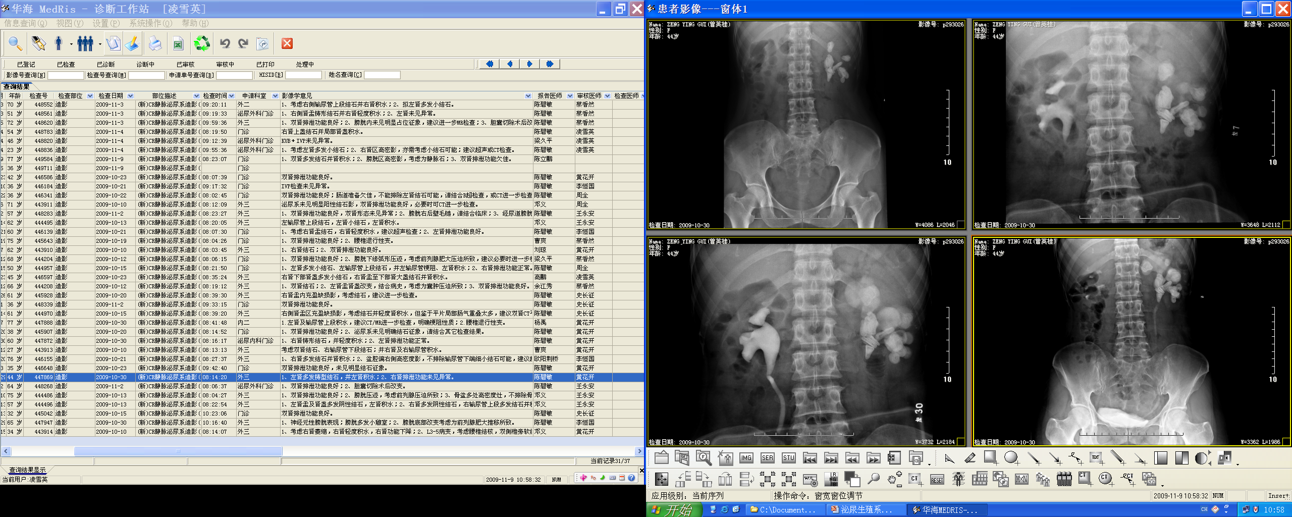
- BNO-IVP (IVU/IVP/excretory urography): contrast IV; depicts renal parenchyma, pelvicalyceal system, ureters, bladder; assesses renal function (filtration/excretion). Phases: 1–3 min nephrogram; 5 min excretory; 15 min pelvicalyceal; 30 min after compression release; 45–60 min full bladder; post-void. Contraindications to compression: suspected stone, acute abdomen, recent abdominal surgery, large mass, aortic aneurysm (use Trendelenburg instead). Largely replaced by CTU.
- Retrograde/antegrade pyelography; retrograde ureterography/cystography/urethrocystography; voiding cystourethrography.
- Ultrasonography: hydronephrosis, kidney stones, bladder mass/stone/diverticulum; first-line in pregnancy/children.
- CT scan: masses, cysts, lesions of kidneys; gold standard in urinary tract stone; tumor staging. Contrast phases: corticomedullary (20–30 s), parenchymal/nephrogenic (60–90 s), excretory (5–10 min).
- CTU (CT urography): illustrates collecting systems, ureters and bladder with IV contrast; has largely replaced IVU; gives anatomical + functional info. Indications: renal calculi, hematuria, characterization of renal mass, staging/follow-up of renal carcinoma, renal vascular anatomy, renal trauma.
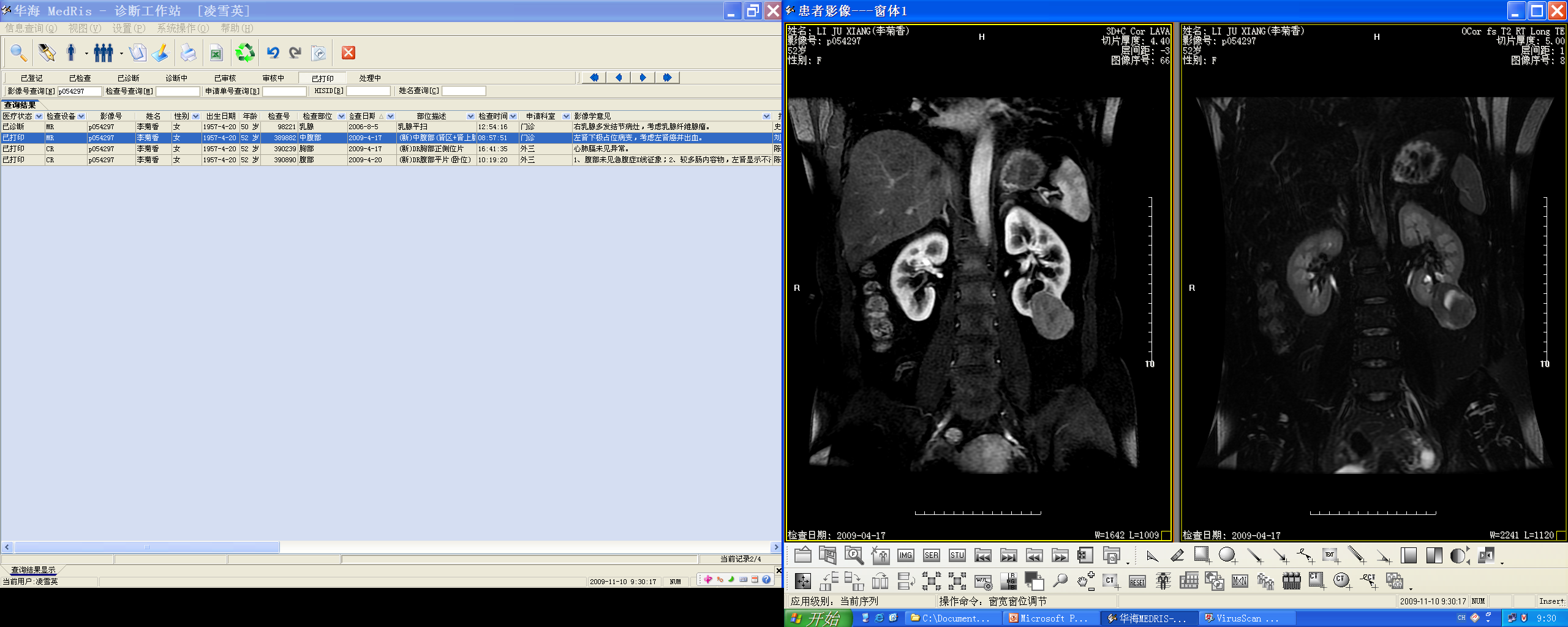
- MRI: staging of bladder and prostatic tumors; best for prostate, uterine and ovary; excellent soft tissue resolution; not sensitive for urinary calculi/calcification.
- MRU: T2-weighted technique like MRCP; evaluation of obstruction, hematuria, congenital anomalies; no contrast, no radiation (pediatric/pregnant).
- Hysterosalpingography (HSG): uterus and fallopian tubes. Indications: infertility (obstruction — can be therapeutic; anomalies e.g. bicornuate uterus), intrauterine pathology (polyps, fibroids, adhesions), post-tubal surgery. Contraindications: pregnancy (perform 7–10 days after menstruation onset), acute PID, active uterine bleeding.
Urinary tract diseases
- Renal/ureteric stones (calculi): 85% visible on plain film; staghorn calculus occupies the collecting system; CT is the gold standard; ureteric stone with hydronephrosis; US shows strong echo + acoustic shadow; IVP shows filling defect/hydronephrosis.
- Hydronephrosis: dilated pelvicalyceal system on US; caused by ureteric stone, stricture (pelviureteric junction narrowing), tumor.
- Renal cyst: simple cyst on US = anechoic, smooth walls, posterior acoustic enhancement; CT = water density, no enhancement; MRI = hypointense T1/hyperintense T2, no enhancement.
- Renal cell carcinoma: solid mass; plain CT = iso/hypodense; contrast CT = heterogeneous enhancement; staging with CT/MRI; tumor may invade the renal vein/IVC (tumor thrombus).
- Bladder carcinoma: bladder mass on US/CT; MRI superior for staging (invasion of wall, perivesical fat, nodes).
Prostate
- Benign prostatic hyperplasia (BPH): involves the transitional zone; major cause of bladder outflow obstruction; PSA elevated but non-specific; MRI: enlarged transitional zone, heterogeneous signal with intact low-signal pseudocapsule.
- Prostate carcinoma: usually arises in the peripheral zone; MRI has higher sensitivity than endorectal US and DRE. T2WI: region of low signal within normally high-signal peripheral zone; DWI/ADC: restricted diffusion (bright on DWI, dark on ADC); DCE: enhancement (can be hard to distinguish from prostatitis/BPH). Extracapsular extension: asymmetry/extension into neurovascular bundles, obliteration of rectoprostatic angle, seminal vesicle involvement (normal seminal vesicles high signal on T2).
Female genital tract
- Uterine fibroids: present in ~25% of women; perimenopausal, family history, Afro-Caribbean; submucosal, subserosal, intramural; MRI best for mapping.
- Ovarian tumors: 5th–7th decades; risk inversely correlated with parity; genetic (BRCA1/2); present late with mass/ascites; classification: epithelial (serous — most common, bilateral in 50%, cystic+solid; mucinous — pseudomyxoma peritonei), sex cord/stromal, germ cell, metastases.
- Cervical carcinoma: MRI (T2WI) for staging — disrupts low-signal fibrous stroma; parametrial invasion (loss of low-signal ring); bladder/rectal invasion; enlarged nodes.
- Endometrial carcinoma: the most common gynecological malignancy; MRI recommended for disease extension; hyperintense on T2WI; myometrial infiltration = interruption of the hypointense junctional zone (>50% = deep invasion).
High-Yield Points
- Key Point: CT is the gold standard for urinary tract stones; CTU for hematuria/renal masses.
- Key Point: IVU phases: 1–3 min nephrogram, 5 min excretory, 15 min pelvicalyceal, 30 min after release, 45–60 min full bladder.
- Key Point: MRI is best for prostate, uterine and ovarian imaging; not sensitive for stones.
- Key Point: Prostate carcinoma = low signal on T2WI in the peripheral zone + restricted diffusion (DWI bright/ADC dark).
- Key Point: BPH = transitional zone, intact pseudocapsule; prostate cancer = peripheral zone.
- Key Point: Endometrial carcinoma is the most common gynecological malignancy; MRI for staging; junctional zone interruption = myometrial invasion.
- Key Point: HSG contraindications: pregnancy, acute PID, active bleeding.
- Key Point: Simple renal cyst = anechoic, smooth wall, posterior enhancement.
LMCHK OSCE Practice(OSCE & LMCHK)
- Painless gross hematuria in an older man → RCC / urothelial carcinoma / stone; initial imaging: CT urography (CTU) or ultrasound + CT; contrast-enhanced CT phases (corticomedullary → nephrogenic → excretory).
- Prostate cancer MRI: "low signal in the peripheral zone on T2, bright on DWI, dark on ADC" — memorize the triple finding.
- Renal colic: CT without contrast (non-contrast CT) is the gold standard for stones — mention stone location and hydronephrosis.
- Ovarian mass: characterize benign vs malignant (solid components, ascites, peritoneal disease → malignant); BRCA history.
- HSG is used for infertility (tubal patency); contraindications matter in OSCE (pregnancy, PID, bleeding).
Topic Summary
GU imaging: US for screening (hydronephrosis, stones, bladder), CT/CTU as the modern workhorse (stones = gold standard; hematuria/masses; phases), MRI for prostate/uterus/ovary staging, MRU for obstruction without radiation, HSG for tubal patency. Diseases: renal stones (85% visible), simple cyst vs RCC (enhancement), bladder carcinoma, BPH (transitional zone) vs prostate cancer (peripheral zone, T2 low + restricted diffusion), fibroids, ovarian tumors (epithelial most common), cervical and endometrial carcinoma staging with MRI (junctional zone).
Illustrations(图解速览)
Case gallery for this chapter — identify the imaging technique, location, features and diagnosis for each: