# Ch05: Pleura, Mediastinum, Hila & Diaphragm(胸膜、纵隔、肺门与膈肌)
Preparatory Mindset
This chapter covers the "extrapulmonary" chest findings: pleural effusion (free and loculated), pleural tumors (metastases and mesothelioma), pneumothorax (including tension), mediastinal masses (hiatal hernia, thymoma, lymphoma, neurogenic tumor), hilar enlargement (pulmonary hypertension vs lymph node enlargement) and the diaphragm. These are favorite exam cases because each has a small, memorable set of imaging features.
Core Concepts
Pleural effusion
- Transudate: congestive heart failure, liver cirrhosis, nephrotic syndrome, peritoneal dialysis, myxoedema, Meigs' syndrome (benign ovarian tumor → ascites + pleural effusion). - Exudate: infections, tumors (lung, breast, lymphoma), pulmonary embolism, vasculitis/autoimmune, empyema thoracis, tuberculous pleuritis, after irradiation, GI disorders (pancreatitis), hemothorax (iatrogenic/traumatic).
- Effusions may lie free in the pleural cavity or become loculated by pleural adhesions (loculation is a particular feature of empyema; a loculated effusion may simulate a lung tumor on CXR).
- Etiology:
- CXR features: homogeneous dense opacity; loss of the costophrenic angle; meniscus sign (higher laterally than medially, concave upper border); loss of hemidiaphragm; no air bronchogram.
- Ultrasound: transonic area between diaphragm and lung; guides thoracentesis.
- CT: homogeneous fluid density, well-defined meniscus-shaped border, compression of adjacent lung; CT is particularly useful for loculated effusions.
- Loculated effusion/empyema: lens-shaped collection with thickened pleura wall; may contain loculated air.
Pleural tumors
- Most common: metastatic carcinomas (e.g. from breast cancer — note absent breast shadow).
- Primary: mesothelioma (asbestos exposure) — lobulated masses + pleural effusion; calcified pleural plaques may coexist.
Pneumothorax
- Most occur in young people with no recognizable lung disease (small peripheral blebs/bullae that burst).
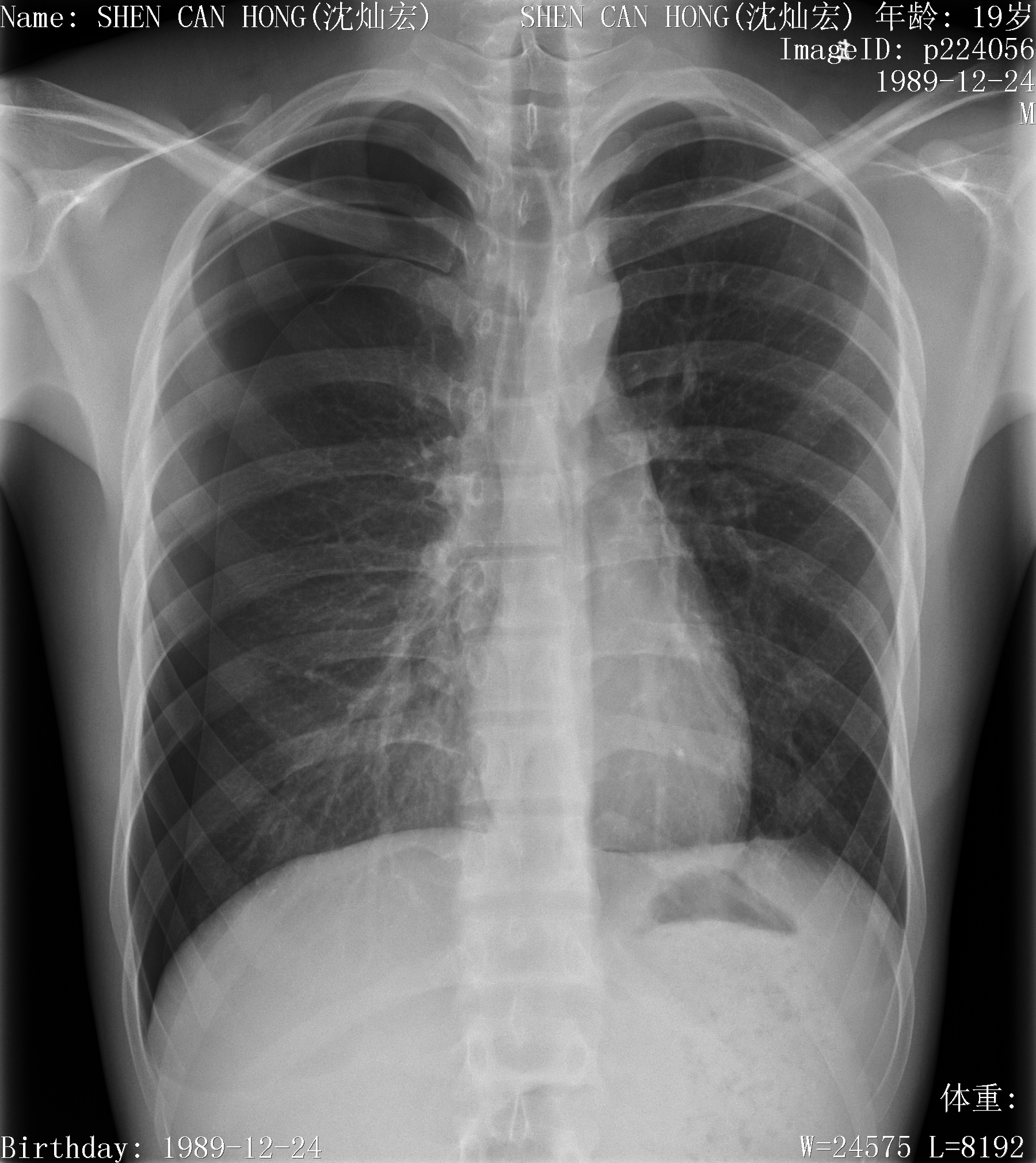
- Imaging features: a line of pleura (lung edge separated by air); no lung markings beyond the lung edge; check for mediastinal shift.
- Tension pneumothorax: mediastinal shift away from the affected side, depressed hemidiaphragm, collapsed lung with increased density — medical emergency.
- Hydropneumothorax/hemopneumothorax/pyopneumothorax: air-fluid levels.
- Pleural edge sign: line approximately parallel to the chest wall; no vessels beyond it.
Mediastinum
- Divided into anterior, middle and posterior divisions for descriptive purposes; masses classified by position.
- CECT of mediastinum distinguishes fat, soft tissue and blood vessels.
- Anterior mediastinal masses: thymoma (large lobulated mass with punctate calcification), lymphoma (extensive mediastinal lymphadenopathy), retrosternal goiter (superior mediastinal mass displacing trachea), teratoma.
- Middle mediastinal masses: lymphadenopathy, bronchogenic cyst (fluid attenuation, no enhancement).
- Posterior mediastinal masses: neurogenic tumors (neurofibroma — MRI: against the spine, not into the canal), hiatal hernia.
- Great vessel disorders: aortic aneurysm (contrast-opacified lumen + unopacified clot lining); aortic dissection (displaced intima separating true and false lumina).
Hilar enlargement
- Vessels (branching pattern, often bilateral) → e.g. severe pulmonary hypertension (bilateral hilar enlargement + bulging pulmonary artery segment).
- Lymph node enlargement → metastases from bronchial carcinoma, lymphoma, infections (TB, histoplasmosis, fungal), sarcoidosis.
- Bilateral hilar adenopathy in sarcoidosis: lobular, dense hila.
- Take-home: CT further evaluates vessel vs mass.
Diaphragm
- Right hemidiaphragm normally up to 2.5 cm higher than the left; at the 6th anterior rib on good inspiration.
- Elevation: ipsilateral lung volume loss, phrenic nerve palsy, subphrenic disease, splenic/liver enlargement.
- Flattening/depression: hyperinflation (emphysema), tension pneumothorax, large effusion.
High-Yield Points
- Key Point: Pleural effusion CXR triad: loss of costophrenic angle + meniscus + homogeneous opacity without air bronchogram.
- Key Point: Transudate vs exudate causes (CHF/cirrhosis/nephrotic vs infection/tumor/PE/autoimmune).
- Key Point: Mesothelioma = asbestos exposure; metastatic pleural tumors are the most common.
- Key Point: Pneumothorax = visceral pleural line with no lung markings beyond; tension = mediastinal shift (emergency).
- Key Point: Mediastinal compartments: anterior = thymoma/lymphoma/goiter; middle = nodes/cyst; posterior = neurogenic tumor/hiatal hernia.
- Key Point: Hilar enlargement: branching = vessels (pulmonary HTN); lobular = lymph nodes (sarcoidosis, lymphoma, mets, TB).
LMCHK OSCE Practice(OSCE & LMCHK)
- Pneumothorax CXR is a classic LMCHK non-interactive station: recognize the visceral pleural line, absent lung markings laterally, and check for mediastinal shift (tension). State: "Right pneumothorax — if there is mediastinal shift to the left with a depressed hemidiaphragm, treat as tension pneumothorax (needle decompression)."
- Effusion vs consolidation: effusion has meniscus + no air bronchogram + loss of costophrenic angle; consolidation has air bronchogram + silhouette sign.
- Mesothelioma: think asbestos exposure history + pleural mass/effusion + calcified plaques.
Topic Summary
Pleural and mediastinal disease is recognized by pattern: effusion = meniscus + obliterated costophrenic angle (CT for loculation); pneumothorax = visceral pleural line with absent lung markings (mediastinal shift = tension, emergency); pleural tumors = metastatic (most common) or mesothelioma (asbestos); mediastinal masses are compartmentalized (anterior: thymoma/lymphoma/goiter; middle: nodes/cyst; posterior: neurogenic/hiatal hernia); hilar enlargement is either vascular (branching, pulmonary HTN) or nodal (lobular, sarcoidosis/lymphoma/mets).
Illustrations(图解速览)
Case gallery for this chapter — identify the imaging technique, location, features and diagnosis for each: