# Ch12: Head & Neck Imaging — Sinuses, Orbits, NPC(头颈部影像)
Preparatory Mindset
Head & neck imaging covers the paranasal sinuses, orbits, mastoid/middle ear, nasopharynx and neck (including salivary glands and thyroid). CT is the preferred technique for sinus anatomy and surgical planning; MRI is better than CT for showing local extent of masses and nodal evaluation. The required master cases: sinusitis, paranasal sinus carcinoma, orbital fractures (blowout/tripod/Le Fort), cavernous hemangioma, retinoblastoma, optic nerve glioma, Graves' disease, mastoiditis, cholesteatoma, and nasopharyngeal carcinoma (NPC).
Core Concepts
Sinuses
- Imaging indicated when simple treatment for inflammatory symptoms fails, or in acute facial trauma.
- Modalities: plain films (Water's view, Caldwell's/PA view, lateral) show mucosal thickening, fluid levels and fractures; CT is the preferred technique; MRI rarely primary; PET/CT, US.
- Acute sinusitis: air-fluid level, total opacification, hyperintense secretions on T2WI.
- Chronic sinusitis: mucosal swelling >5 mm, lumen stenosis + sclerosis, hyperattenuation lesion on NCCT, hypointense secretion on T1/T2.
- Osteomeatal complex (OMC): middle meatus drainage pathway — key to understanding obstruction.
- Paranasal sinus carcinoma: maxillary sinus (80%), nasal cavity (10%), ethmoid (5–6%); squamous cell carcinoma (80%); >40 y in 95%, M:F 2:1; asymmetry of face; bony destruction (90%) predominates over expansion; cervical LN metastases 10–18%.
- Mucocele: mucus-filled sinus, expands and thins walls.
Orbits
- Modalities: CT (coronal cuts for fracture) and MRI; CT and MRI clearly demonstrate anatomy and distinguish intra- vs extra-orbital masses and thyroid eye disease; relationship to the optic nerve is determined.
- Blowout fracture: involves the floor of the orbit (or roof of maxillary sinus); anterior rim usually intact; inferior herniation of orbital fat or entrapment of the inferior rectus; diplopia on upward gaze; CT with coronal reconstructions; crescentic soft tissue mass in the roof of the antrum.
- Tripod fracture: three sites — frontozygomatic suture, zygomatic arch, maxilla; involves the anterior rim of the orbit; mobile, often slightly depressed zygoma.
- Le Fort fractures: type I, II, III (facial disjunction).
- Cavernous hemangioma: most common adult orbital mass (12–15%); slowly progressive unilateral proptosis, diplopia, diminished visual acuity; well-circumscribed intraconal round mass; MRI low T1/high T2; CT tiny calcification; variable enhancement.
- Retinoblastoma: rare malignant congenital intraocular tumor from primitive photoreceptor cells/retina; 98% in children <5 y; leukocoria; 60% unilateral, 40% bilateral; solid retrolental hyperdense mass with punctate/nodular calcification (most common cause of orbital calcifications); exophytic vs endophytic vs diffuse forms; iso/mild hyperintense T1, hypointense T2, moderate-marked enhancement.
- Optic nerve glioma (= juvenile pilocytic astrocytoma): most common cause of optic nerve enlargement; 80% in first decade; 15% with neurofibromatosis; ipsilateral optic canal enlargement >3 mm; MRI more sensitive (iso T1, hyper T2, Gd enhancement).
- Graves' disease (thyroid ophthalmopathy): enlargement of extraocular muscles — bilateral 70–85%; I'M Slow (Inferior > Medial > Superior > Lateral); proptosis (globe >21 mm anterior to interzygomatic line); sparing of tendinous insertion = coke bottle sign; high T2 signal in enlarged muscles (acute edema).
Mastoid and middle ear
- Modalities: Schüller view, Mayer's view, high-resolution CT (HRCT), MRI.
- Mastoiditis: cloudy mastoid cells, thickened trabeculae, complete sclerosis; ossicular erosion (long process of incus, handle of malleus).
- Cholesteatoma (= keratoma): keratin debris causing bone destruction by pressure and demineralizing enzymes. Primary (congenital, 2%) vs secondary (acquired inflammatory, 98%). Imaging: soft tissue mass in the middle ear, no contrast enhancement, poorly aerated mastoid, ossicular destruction, bony erosion of facial nerve canal and semicircular canals; MR to rule out recurrent cholesteatoma.
Nasopharynx and neck
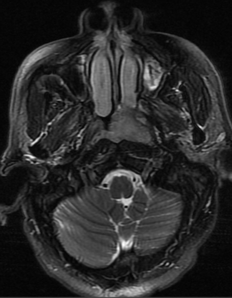
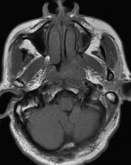
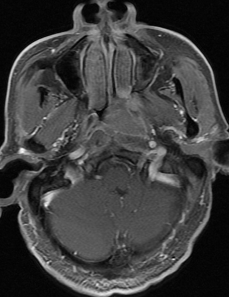
- Imaging: nasopharyngeal wall thickening, shallow or disappeared pharyngeal recess, narrowed pharyngeal cavity; invasion of nasal cavity/masticator space/sinuses; base of skull destruction; MR better than CT for local extent + nodal evaluation; imaging required to detect skull base spread, perineural extension and lymphadenopathy.
- Normal axial CT of NP: symmetry, regular soft tissue layer, well-defined lateral wall structures (pharyngeal recess/Rosenmuller fossa, pharyngeal opening of auditory tube), well-defined fat planes.
- Nasopharyngeal carcinoma (NPC): the most common malignant tumor of the nasopharynx; a squamous cell carcinoma that usually presents late with local invasion and necrotic lymph node metastases; Chinese population predominant, mean age 40; unilateral nasal obstruction, history of chronic sinusitis.
- Laryngeal carcinoma: best examined with CT (less movement artifact than MR); targeted MRI for soft tissue; tumor destroys vocal cords, invades thyroid cartilage; nodal metastases.
- Salivary glands: pleomorphic adenoma (parotid) — high signal on T2-weighted MRI; sialography demonstrates duct strictures/stones.
- Thyroid: nodular goiter, thyroid carcinoma; tracheal compression suggests malignant mass; radioiodine detects metastases before ablation; parathyroid adenomas — Tc-99m pertechnetate washes out more slowly from parathyroid tissue.
Cervical lymph node assessment
- Up to 300 nodes, 3 mm–3 cm normal; most within 1–2 cm.
- Criteria of malignancy: size, shape, necrosis, extracapsular spread, vascular flow, number, calcification.
High-Yield Points
- Key Point: CT = sinus anatomy/surgery planning + bone destruction; MRI = local extent + nodes (NPC).
- Key Point: Acute sinusitis = air-fluid level/opacification; chronic = mucosal thickening >5 mm + sclerosis.
- Key Point: Blowout fracture = orbital floor + inferior rectus entrapment + diplopia on upgaze; CT coronal.
- Key Point: Retinoblastoma = child <5 y + calcified intraocular mass (most common orbital calcification).
- Key Point: Graves' = extraocular muscle enlargement sparing tendons (coke bottle sign); Inferior > Medial > Superior > Lateral (I'M Slow).
- Key Point: Cholesteatoma = non-enhancing middle-ear soft tissue mass + ossicular/bone erosion.
- Key Point: NPC = pharyngeal recess obliteration + skull base destruction + necrotic cervical nodes; Chinese, mean 40 y.
- Key Point: Malignant neck node criteria: size, shape, necrosis, extracapsular spread, vascularity.
LMCHK OSCE Practice(OSCE & LMCHK)
- NPC is a listed LMCHK common case (HNS): unilateral nasal obstruction + epistaxis in a Chinese adult; imaging shows nasopharyngeal mass obliterating the pharyngeal recess with skull-base erosion and necrotic cervical nodes; MRI for extent; treatment: radiotherapy ± chemo.
- Blowout fracture: history of blow to eye → coronal CT shows orbital floor fracture with herniated orbital fat / entrapped inferior rectus; diplopia on upgaze.
- Retinoblastoma: child with leukocoria → CT shows calcified intraocular mass; MRI to assess optic nerve/extraocular extension.
- Sinusitis: acute (air-fluid level) vs chronic (mucosal thickening + sclerosis); CT for surgical planning.
- Laryngeal cancer: CT showing vocal cord destruction + thyroid cartilage invasion + nodal metastasis.
Topic Summary
Head & neck imaging: sinuses (CT preferred; acute = air-fluid level, chronic = mucosal thickening, carcinoma = bone destruction), orbits (CT for fractures — blowout/tripod/Le Fort; MRI for masses — cavernous hemangioma, retinoblastoma with calcification, optic glioma, Graves' with I'M Slow muscle enlargement), mastoid/middle ear (HRCT; mastoiditis; cholesteatoma = non-enhancing erosive mass), and nasopharynx/neck (NPC with skull-base destruction + necrotic nodes; laryngeal carcinoma; salivary/thyroid; malignant node criteria). MRI wins for soft-tissue extent and nodes; CT wins for bone and sinus anatomy.
Illustrations(图解速览)
Case gallery for this chapter — identify the imaging technique, location, features and diagnosis for each: