Preparatory Mindset
Abdominal pain, diarrhea, hematemesis, and hematochezia are the core GI symptoms. The #1 rule: rule out the surgical abdomen and life-threatening causes (ruptured AAA, perforated viscus, mesenteric ischemia, ruptured ectopic pregnancy) before anything else. Understand the three pain mechanisms (visceral, somatic, referred), the embryologic localization (foregut/midgut/hindgut), and the classic per-region differentials. For bleeding, always quantify and differentiate upper (hematemesis/melena) from lower (hematochezia).
Illustrations(图解速览)
Core Concepts — Abdominal Pain (腹痛)
Etiology — Acute vs Chronic
Acute (<1 week):
- Acute organ inflammation: acute gastritis/enteritis, acute pancreatitis, acute hemorrhagic necrotic enteritis, acute cholecystitis, acute appendicitis
- Stretching/distention: intestinal obstruction, bile duct blocked by gallstones
- Twisting/rupture: volvulus, mesenteric/omental torsion, hepatic/splenic rupture, ruptured ectopic pregnancy
- Peritoneal inflammation: GI perforation, spontaneous peritonitis
- Loss of blood supply: ischemic bowel disease, portal vein thrombosis
- Abdominal wall: injury, herpes zoster
- Referred from thorax: pneumonia, pulmonary infarction, angina, MI, acute pericarditis
- Systemic: Henoch–Schönlein purpura, DKA, uremia, lead poisoning, porphyria
Chronic:
- Chronic inflammation: reflux esophagitis, chronic gastritis/cholecystitis/pancreatitis, TB peritonitis, UC, Crohn's
- Hollow viscera tension change: GI spasm
- Peptic ulcer
- Chronic twisting/block (volvulus)
- Organ swelling (hepatitis hepatomegaly)
- Poisoning/metabolic (uremia, lead poisoning)
- Tumor
- GI neurosis
Pathogenesis — 3 Pain Types
| Type | Mechanism | Features |
|---|---|---|
| Visceral pain | Autonomic nerves in visceral peritoneum; sympathetic route | Poorly localized, intermittent/crampy/colicky, with vagal symptoms (nausea, vomiting, diaphoresis) |
| Somatic pain | Parietal peritoneum irritation; peripheral nerves | Precisely localized, intense/constant, guarding/rigidity, worse with cough/position change |
| Referred pain | Same segmental innervation projected to superficial region | Pain at a distance from source (MI → neck/shoulders/back; cholecystitis → right scapula; renal colic → perineum; appendicitis → RLQ after periumbilical onset) |
Embryologic Localization (Foregut/Midgut/Hindgut)
- Foregut (stomach, pancreas, duodenum, liver, gallbladder) → epigastric
- Midgut (small bowel, proximal colon, spleen) → periumbilical
- Hindgut (distal colon) → suprapubic/hypogastric
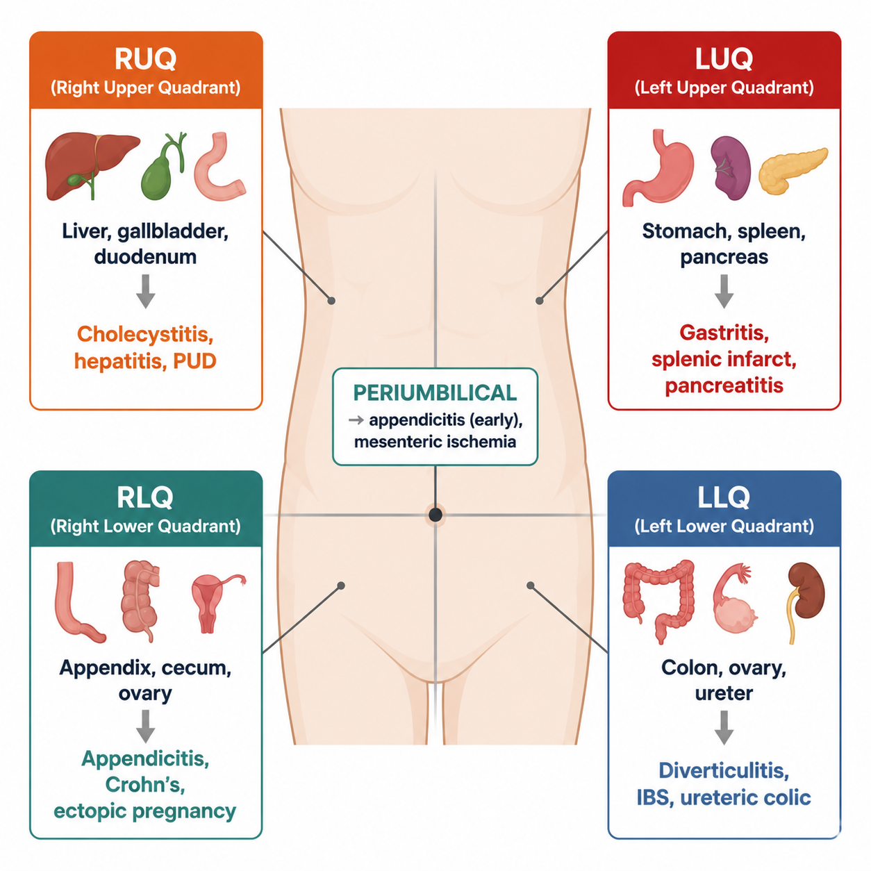
Pain by Location — Key Differentials
| Region | Likely cause | Key features |
|---|---|---|
| RUQ | Cholecystitis, cholangitis, hepatitis | Biliary colic steady >1h after meals; Charcot's triad (RUQ pain + jaundice + fever) = cholangitis (emergency); hepatitis = tender enlarged liver + malaise + jaundice |
| Epigastrium | Pancreatitis, PUD, AAA dissection | Pancreatitis: post-heavy-meal/alcohol, constant mid-epigastric, radiates to back, ↑amylase/lipase; PUD: burning, food/NSAID/H. pylori related; dissection: tearing, midline back, hypotensive, pulsatile mass |
| LUQ | Peptic ulcer, splenic pathology, dissection | — |
| RLQ | Appendicitis | Periumbilical → RLQ migration, McBurney tenderness, fever, leukocytosis |
| LLQ | Diverticulitis, ovarian cysts | Cramping steady; avoid colonoscopy/barium enema in acute diverticulitis |
| Suprapubic | Pelvic (bladder, uterus, ovary, ectopic pregnancy) | Always consider pelvic causes in females with acute abdomen |
Immediate life-threats requiring rapid surgery
Ruptured abdominal aortic aneurysm; perforated stomach/intestine (free air); mesenteric ischemia; ruptured ectopic pregnancy.
Core Concepts — Diarrhea (腹泻)
Definition & Classification
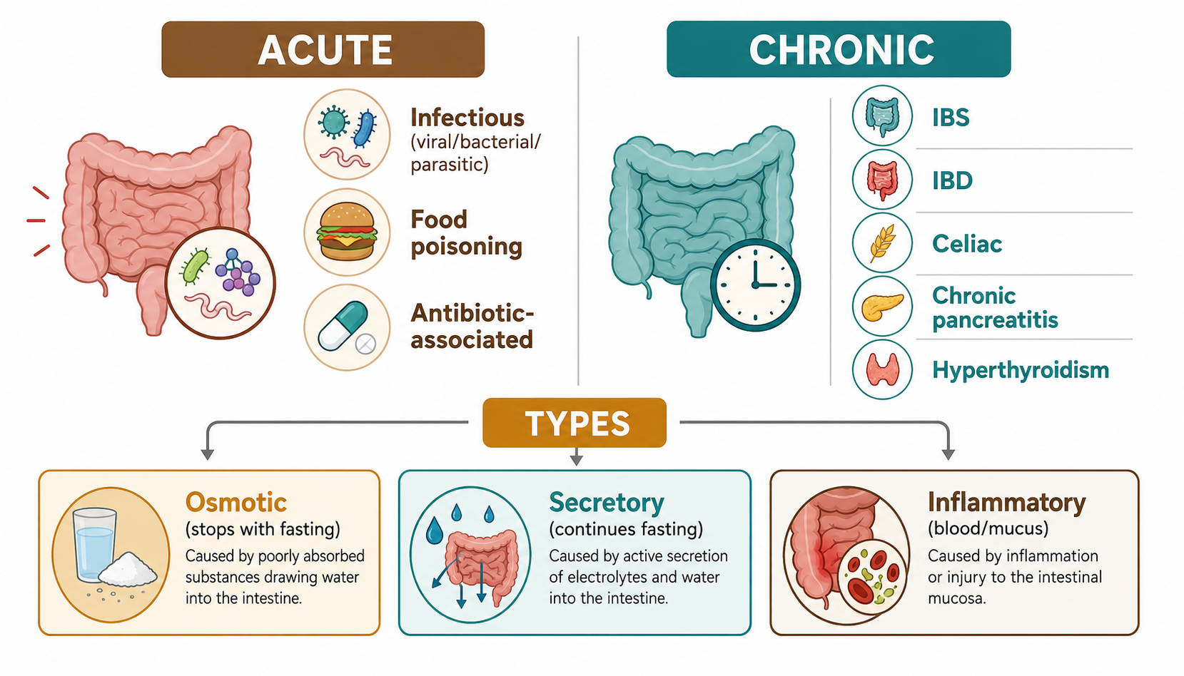
- Diarrhea = increased stool frequency (>3/day) with loose/watery stools, or increased volume (>200 g/day)
- Acute (<3 weeks) vs Chronic (>3–4 weeks)
- Secretory: large volume, persists with fasting, normal osmotic gap — cholera, VIPoma, laxative abuse
- Osmotic: stops with fasting, high osmotic gap — lactose intolerance, osmotic laxatives (Mg)
- Inflammatory/exudative: blood, pus, mucus — IBD, infectious colitis
- Steatorrhea: fatty, greasy, foul — malabsorption, pancreatic insufficiency
Causes
- Acute: infection (bacterial, viral, parasitic), food poisoning, drugs (antibiotics), ischemic colitis
- Chronic: IBS, IBD (UC/Crohn's), malabsorption (celiac, pancreatic), endocrine (hyperthyroidism, DM), colon cancer, laxative abuse
History-taking points
Onset, frequency, volume, consistency, blood/mucus, nocturnal diarrhea (organic > functional), relationship to food, weight loss, fever, antibiotics, travel, family history, tenesmus.
Core Concepts — Hematemesis & Melena (呕血与黑便) / Hematochezia (便血)
Definitions
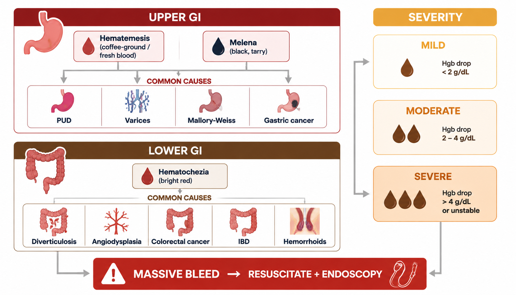
- Hematemesis (呕血): vomiting of blood — upper GI bleeding (above the ligament of Treitz)
- Melena (黑便/tarry stool): black, sticky stools from digested blood — usually upper GI, ≥50–100 ml blood
- Hematochezia (便血): bright red blood per rectum — usually lower GI or massive upper GI bleed
Etiology — Upper GI Bleeding
- Peptic ulcer (most common) — H. pylori, NSAIDs
- Esophageal/gastric varices (portal hypertension, cirrhosis)
- Mallory-Weiss tear (post-retching)
- Erosive gastritis/esophagitis
- Tumors, angiodysplasia, Dieulafoy lesion
- Systemic: coagulopathy, DIC, leukemia
Lower GI Bleeding
- Hemorrhoids (common), anal fissure, diverticulosis, angiodysplasia, IBD, colon cancer, ischemic colitis, infectious colitis
Clinical Evaluation
- Estimate amount: mild <500 ml; moderate 500–1000 ml; severe >1000 ml or signs of shock
- Assess hemodynamic status first: BP, HR, orthostatic change, urine output, signs of shock
- Determine site: hematemesis/melena → upper (EGD); hematochezia → lower (colonoscopy) but massive upper bleed can present as hematochezia
- History: prior ulcer, NSAIDs/alcohol, liver disease, anticoagulants, family history (coagulopathy, polyposis)
- Exam: pallor, tachycardia, hypotension, stigmata of liver disease (spider angioma, caput medusa), melena on DRE
High-Yield Points
- Surgical abdomen red flags: rigid abdomen, rebound tenderness, free air, hypotension + pulsatile mass (AAA), periumbilical→RLQ migration (appendicitis)
- Visceral pain = poorly localized + vagal symptoms; somatic = localized + guarding; referred = same dermatome
- Foregut→epigastric, midgut→periumbilical, hindgut→suprapubic
- Charcot's triad (RUQ pain + jaundice + fever) = ascending cholangitis — emergency
- Pancreatitis: epigastric → back, after alcohol/heavy meal; ↑amylase/lipase
- Melena = ≥50–100 ml upper GI blood; hematemesis = above ligament of Treitz
- GI bleed management: resuscitation first, then locate (EGD/colonoscopy), then treat (PPI, endoscopic therapy, variceal banding/TIPS)
- Cullen's sign (periumbilical bruising) & Grey-Turner's sign (flank bruising) = retroperitoneal bleeding (severe pancreatitis, trauma, ectopic rupture)
LMCHK OSCE Practice
- Abdominal pain history: onset (sudden/gradual), site & radiation, character (colicky/constant/burning/tearing), severity, aggravating/relieving (food, posture, defecation), associated (nausea/vomiting, fever, bleeding, jaundice, weight loss), past surgery, alcohol/NSAIDs, pregnancy test in women of childbearing age!
- GI bleed: ask about hematemesis vs melena vs hematochezia; prior ulcer/varices; NSAIDs/aspirin/anticoagulants; check vitals + DRE (melena)
- Abdominal exam common cases (LMCHK): liver, kidney, hematological diseases — assess hepatosplenomegaly, ascites (shifting dullness, fluid thrill), and surgical scars
- Acute abdomen: peritonism, guarding, rebound — urgent surgical referral
Topic Summary
Abdominal pain: classify acute vs chronic; understand visceral/somatic/referred mechanisms and foregut-midgut-hindgut localization; build per-region differentials (RUQ=biliary, epigastric=pancreas/PUD, RLQ=appendicitis, LLQ=diverticulitis, suprapubic=pelvic). Diarrhea: secretory vs osmotic vs inflammatory. GI bleeding: distinguish hematemesis (upper) / melena (upper, ≥50 ml) / hematochezia (lower or massive upper); resuscitate first, then localize and treat.