Preparatory Mindset
Serous membrane effusions (pleural, pericardial, peritoneal) accumulate when fluid formation exceeds lymphatic drainage. The central exam question is always: transudate or exudate? Master Light's criteria, the SAAG for ascites, the key biochemical discriminators (protein, LDH, glucose, ADA), and the procedures — thoracentesis and abdominocentesis — with their indications, contraindications, and complications. This is a high-yield, heavily tested topic with clear tables.
Illustrations(图解速览)
Core Concepts — Serous Fluid Formation
Anatomy & normal physiology
- Serous cavities: pleural (lungs), pericardial (heart), peritoneal (abdomen/pelvis)
- Lined by two mesothelial membranes: parietal (lines cavity wall) + visceral (covers organ)
- Serous fluid = ultrafiltrate of plasma — formation depends on hydrostatic vs colloid (oncotic) pressure balance; normally small amount (lubrication), production = reabsorption
- Effusion develops when formation > lymphatic removal
Transudate vs Exudate — mechanism
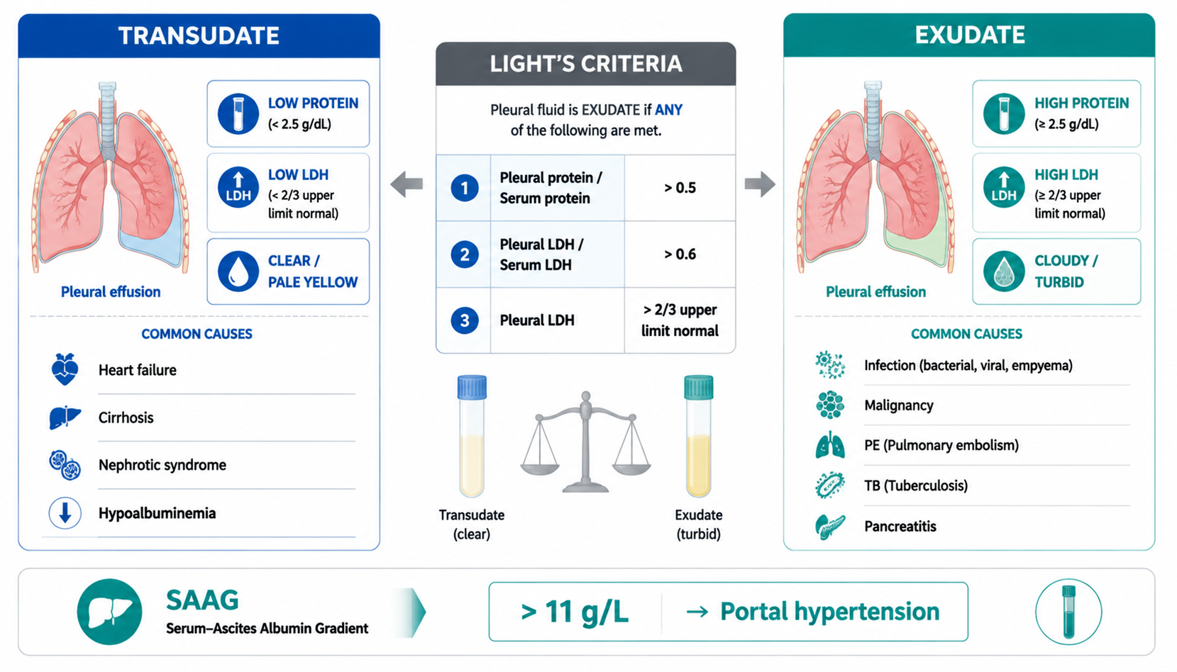
- Transudate: altered systemic factors — ↑hydrostatic pressure (heart failure), ↓oncotic pressure (nephrotic syndrome, cirrhosis, hypoalbuminemia) — pleura itself normal
- Exudate: altered pleural surface — inflammation/tumor ↑ capillary permeability or ↓lymphatic drainage (pleurisy, malignancy, PE, SLE, TB)
Core Concepts — Pleural Fluid Analysis (胸水)
Light's criteria (exudate if any one) — MUST KNOW
- Pleural fluid protein / serum protein >0.5
- Pleural fluid LDH / serum LDH >0.6
- Pleural fluid LDH >2/3 upper limit of normal serum (or >200 IU/L per some sources)
Transudate vs Exudate — comparison table
| Feature | Transudate | Exudate |
|---|---|---|
| Appearance | Light yellow, clear | Yellow, turbid, purulent, bloody |
| Specific gravity | <1.018 | >1.018 |
| Coagulability | Does not clot | Clots |
| Rivalta test | Negative | Positive |
| Protein | <30 g/L | >30 g/L |
| PF/Serum protein | <0.5 | >0.5 |
| LDH | <200 IU/L | >200 IU/L |
| PF/Serum LDH | <0.6 | >0.6 |
| WBC | <100×10⁶/L (lymphocytes, mesothelial) | >500×10⁶/L (PMN = acute inflammation; lymphocytes = TB/tumor; eosinophils = parasite/CTD) |
| Glucose | Similar to serum | ↓ (pyothorax, SLE, TB, malignancy) |
| pH | ~7.6 | <7.3 (infection, CTD, RA, TB); <7.0 = pyothorax/esophageal rupture; >7.35 = SLE/malignancy |
Specific markers — HIGH YIELD
| Marker | Meaning |
|---|---|
| ADA >45 U/L | Tuberculous pleurisy |
| LDH >500 IU/L | Malignant or infective |
| CEA >20 µg/L, PF/Serum >1 | Malignant effusion |
| Amylase ↑ | Pancreatitis, esophageal rupture, tumor |
| Glucose <3.3 mmol/L | Pyothorax, SLE, TB, rheumatoid, malignancy |
| RBC >5000×10⁶/L | Malignant, TB; >10000 (hemothorax) — trauma, tumor, PE |
| Triglyceride >1.21 mmol/L, milky | Chylothorax |
| Chocolate color | Amebic (look for trophozoites) |
| Lupus cells | SLE |
| Cytology | Malignant cells |
| Smear/culture | MTB positive ~20% in TB pleurisy |
- pH <7.0 = pyothorax or esophageal rupture (consider chest tube); pH <7.3 + glucose <3.3 = complicated parapneumonic (drainage indication)
- Exudate causes: pneumonia (parapneumonic), TB, malignancy (metastatic, mesothelioma), PE, SLE/RA, pancreatitis
- Transudate causes: CHF (most common overall), cirrhosis/hepatic hydrothorax, nephrotic syndrome, hypoalbuminemia, myxedema, constrictive pericarditis, SVC obstruction
Core Concepts — Ascites Analysis (腹水)
SAAG (serum-ascites albumin gradient) — MUST KNOW
- SAAG = serum albumin − ascitic albumin
- High SAAG (≥11 g/L): portal hypertension — cirrhosis, heart failure, Budd-Chiari, massive liver metastases
- Low SAAG (<11 g/L): non-portal — peritoneal carcinomatosis, TB peritonitis, nephrotic syndrome (hypoalbuminemia), pancreatic ascites, biliary ascites
- Note: total protein is NOT reliable for ascites (SAAG replaces it); serum-ascites protein gradient useful in some contexts
Ascitic fluid tests
- Cell count/differential: PMN ≥250/mm³ = spontaneous bacterial peritonitis (SBP) — culture + antibiotics
- Protein, albumin (SAAG), LDH, glucose, ADA (TB), amylase (pancreatic), cytology (malignancy), Gram stain/culture, triglyceride (chylous)
Causes of ascites
- Portal hypertension (cirrhosis, HF, Budd-Chiari, constrictive pericarditis)
- Peritoneal disease (carcinomatosis, TB, SBP, pancreatic)
- Hypoalbuminemia (nephrotic, malnutrition)
Core Concepts — Thoracentesis (胸腔穿刺术)
Indications
- Diagnostic: any pleural effusion of unknown cause (clinically significant — >10 mm on USS/lateral decubitus)
- Therapeutic: relieve dyspnea from pneumothorax/large effusion; drain + rinse pyothorax; instill drugs (antibiotics, chemotherapy)
Contraindications (relative — no absolute)
Coagulation disorder, anticoagulant therapy, chest wall cellulitis at site, uncertain fluid location, minimal fluid, uncontrolled cough, altered chest wall anatomy, uncooperative patient
Preparation
- Informed consent; comfort patient; sterile technique
- Equipment: lidocaine 1–2% (10 ml), gauze, 5-ml syringe, antiseptic, drapes, gloves, thoracentesis bag (needle + catheter), 3-way stopcock, 30–50 ml syringe, dressings, collection tubes (red/purple top, blood culture), vacuum bottle (large volume), USS (if guided), epinephrine (rescue)
Positioning & site
- Sitting with arms supported on bedside table + footstool; lateral recumbent; semi-Fowler's
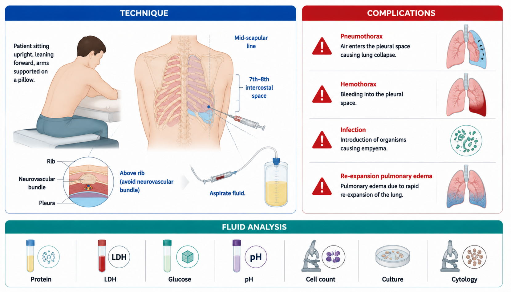
- Pleural effusion: 7th–9th ICS, scapular line or posterior axillary line (confirmed by dullness/↓fremitus + USS/CXR)
- Pneumothorax: 2nd–3rd ICS, midclavicular line
- Insert on superior edge of rib — the intercostal neurovascular bundle runs along the lower edge of each rib
- Be careful of liver (right, up to 5th ICS on expiration) and spleen (left, up to 9th ICS)
Procedure & amounts
- Sterile prep + drape; anesthetize skin with lidocaine (wheal), advance with aspiration, inject 0.1–0.2 ml lidocaine every 1–2 mm
- Insert 22-gauge needle attached to 30-ml syringe, above the rib, aspirate while advancing; a "pop" may be felt entering the pleura; note depth
- Diagnostic: 50–100 ml; Therapeutic: first time ≤600 ml, then ≤1000 ml per session; Pneumothorax: ≤1000 ml at a time
- If >500 ml withdrawn, monitor symptoms/BP — stop if chest pain, dyspnea, or hypotension
- Remove needle, sterile dressing; process fluid (transudate vs exudate)
Complications
- Pneumothorax (most common — including tension pneumothorax: cyanosis, dyspnea, sweating, hypotension → urgent decompression), bleeding/hemothorax, re-expansion pulmonary edema (large rapid drainage), infection, organ puncture (liver/spleen/diaphragm), vasovagal reaction
Core Concepts — Abdominocentesis (腹腔穿刺术)
Indications
- Diagnostic: new-onset ascites of unknown etiology; diagnose SBP, TB, metastatic cancer, blood in peritoneal cavity (trauma)
- Therapeutic: relieve pressure (large ascites with severe distension, chest tightness, SOB, oliguria); intraperitoneal drugs/chemotherapy (hyperthermic perfusion); ascites concentration recycling; artificial pneumoperitoneum
Contraindications
- Absolute: acute abdomen requiring surgery
- Relative: PT >21 s / INR >1.6 / platelets <50,000 (increased bleeding); late pregnancy, ovarian cyst, hydatid, distended bladder, abdominal wall cellulitis, severe distended bowel, severe adhesions, early hepatic encephalopathy/restless patient
Procedure
- Consent + education; empty bladder; supine, head elevated 20–30°
- Sites: ①outer 1/3 of line between umbilicus and ASIS; ②umbilicus horizontal line × anterior/mid-axillary (lateral decubitus); ③1 cm above umbilicus-pubis midpoint, 1.5 cm lateral; ④USS-guided; avoid scars
- Sterile prep (15 cm diameter, 3×), gloves; lidocaine infiltration down to peritoneum (aspirate periodically)
- Z-tracking (advance 1–2 cm after piercing skin, then redirect) to prevent fluid leak
- Slow advance with aspiration; peritoneal "give" felt; diagnostic = 50 ml
- Large-volume: direct drainage to vacuum containers; limit ≤3 L (cirrhosis); albumin 1–2 units after 4–5 L
- Remove, sterilize, pressure dressing; supine 2–4 h
Complications & management
| Complication | Management |
|---|---|
| Fluid not flowing | Adjust depth/site (USS) |
| Feculent tap | Withdraw, new site, observe 24 h for peritonitis |
| Bloody tap | Check coagulation before; withdraw; monitor |
| Hypotension after large-volume paracentesis | IV saline bolus, Trendelenburg, albumin after >4–5 L |
| Infection, bowel perforation | Antibiotics, surgical review |
High-Yield Points
- Light's criteria (exudate if ≥1): PF/Serum protein >0.5; PF/Serum LDH >0.6; PF LDH >2/3 upper normal
- Transudate: protein <30, LDH <200, SG <1.018, Rivalta negative — CHF/cirrhosis/nephrotic
- Exudate: protein >30, LDH >200, Rivalta positive — infection/malignancy/TB/PE/SLE
- ADA >45 U/L = TB pleurisy; CEA >20 = malignant; glucose <3.3 = pyothorax/SLE/TB/RA; chylous (TRIG >1.21) = chylothorax
- pH <7.0 = pyothorax/esophageal rupture (chest tube)
- SAAG ≥11 = portal hypertension ascites; <11 = carcinomatosis/TB/nephrotic
- SBP: ascitic PMN ≥250/mm³ → antibiotics (no need for culture wait)
- Thoracentesis: above the rib (neurovascular bundle below); effusion 7th–9th ICS scapular/posterior axillary; pneumothorax 2nd–3rd ICS MCL
- Therapeutic thoracentesis: first ≤600 ml then ≤1000 ml; pneumothorax ≤1000 ml — risk re-expansion pulmonary edema
- Tension pneumothorax after thoracentesis: cyanosis + dyspnea + hypotension → immediate decompression
- Abdominocentesis: Z-tracking, empty bladder, ≤3 L (albumin after 4–5 L), SBP diagnosis
- Pericardial effusion/tamponade: fluid in pericardial sac compresses heart — Beck's triad (hypotension, muffled sounds, JVD)
LMCHK OSCE Practice
- Pleural effusion OSCE: state findings (↓expansion, ↓fremitus, stony dullness, ↓breath sounds, ↓vocal resonance, trachea away if massive) → propose diagnostic thoracentesis + USS-guided; send fluid for protein, LDH, glucose, ADA, cytology, culture; classify transudate/exudate (Light's criteria)
- Ascites exam: shifting dullness + fluid thrill → diagnostic paracentesis → SAAG + PMN count (SBP screen) + cytology + ADA (TB)
- Comment: "This is a transudative effusion (protein 25 g/L, LDH 150, PF/S <0.5) — most likely heart failure; I would treat the underlying cause and consider diuretics"
- Exudative: "consistent with infection or malignancy — I would arrange CXR/CT, sputum AFB, and pleural biopsy if TB suspected"
- Procedure stations: state indications, contraindications, consent, positioning, site, volumes, complications before doing the technique
- Empyema/parapneumonic with pH <7.2 or glucose <3.3 → chest tube drainage, not repeated taps
Topic Summary
Serous effusions: transudate (systemic — HF/cirrhosis/nephrotic) vs exudate (surface — infection/malignancy/TB/PE); classify with Light's criteria + protein/LDH/SG/Rivalta. High-yield markers: ADA (TB), CEA (malignant), glucose/pH (complicated parapneumonic), chylothorax (TRIG). Ascites: SAAG (≥11 portal HTN) + PMN ≥250 = SBP. Thoracentesis: above rib, effusion 7th–9th ICS, pneumothorax 2nd–3rd ICS, ≤600 ml first then 1000 ml, tension pneumothorax complication. Abdominocentesis: Z-tracking, ≤3 L, albumin after large volume. Master the tables — this chapter is guaranteed exam and OSCE material.