Preparatory Mindset
The cardiovascular examination is the most demanding system exam — inspection, palpation, percussion, and above all auscultation. Master the 5 auscultatory valve areas, the normal S1/S2, extra heart sounds (gallops, opening snap, clicks), and the murmur characterization (location, timing, quality, intensity, radiation) (18CM quiz tested) (18CM quiz tested). Then map murmurs to valve lesions (AS, AR, MS, MR, TR) — the single most-tested content in both school exams and LMCHK CVS short cases.
Illustrations(图解速览)
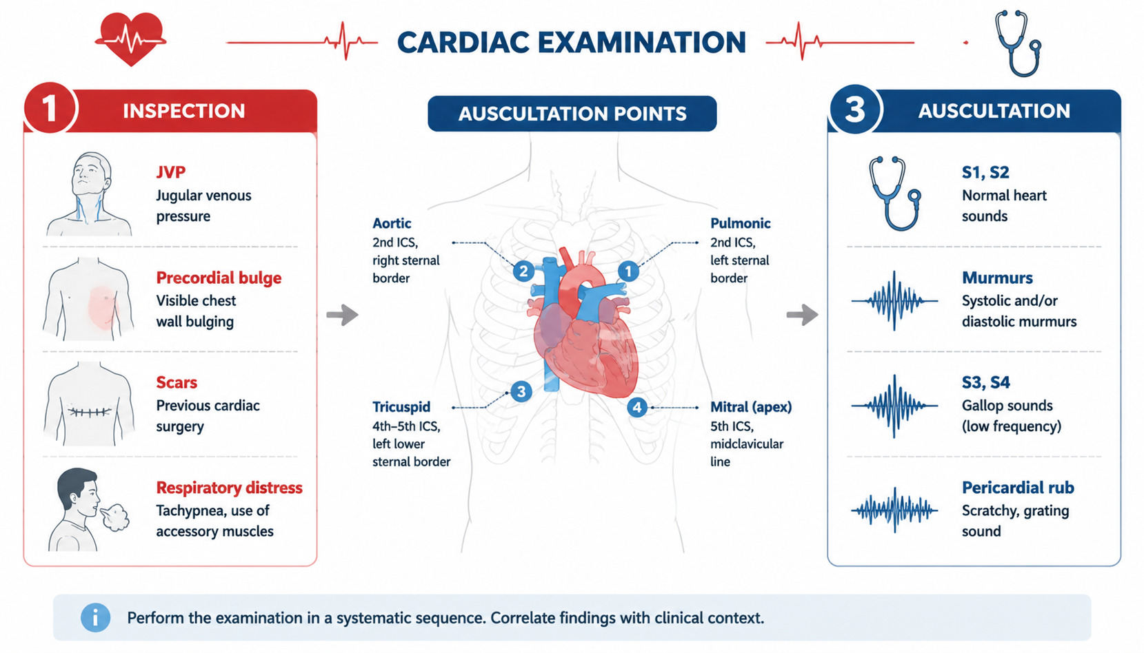
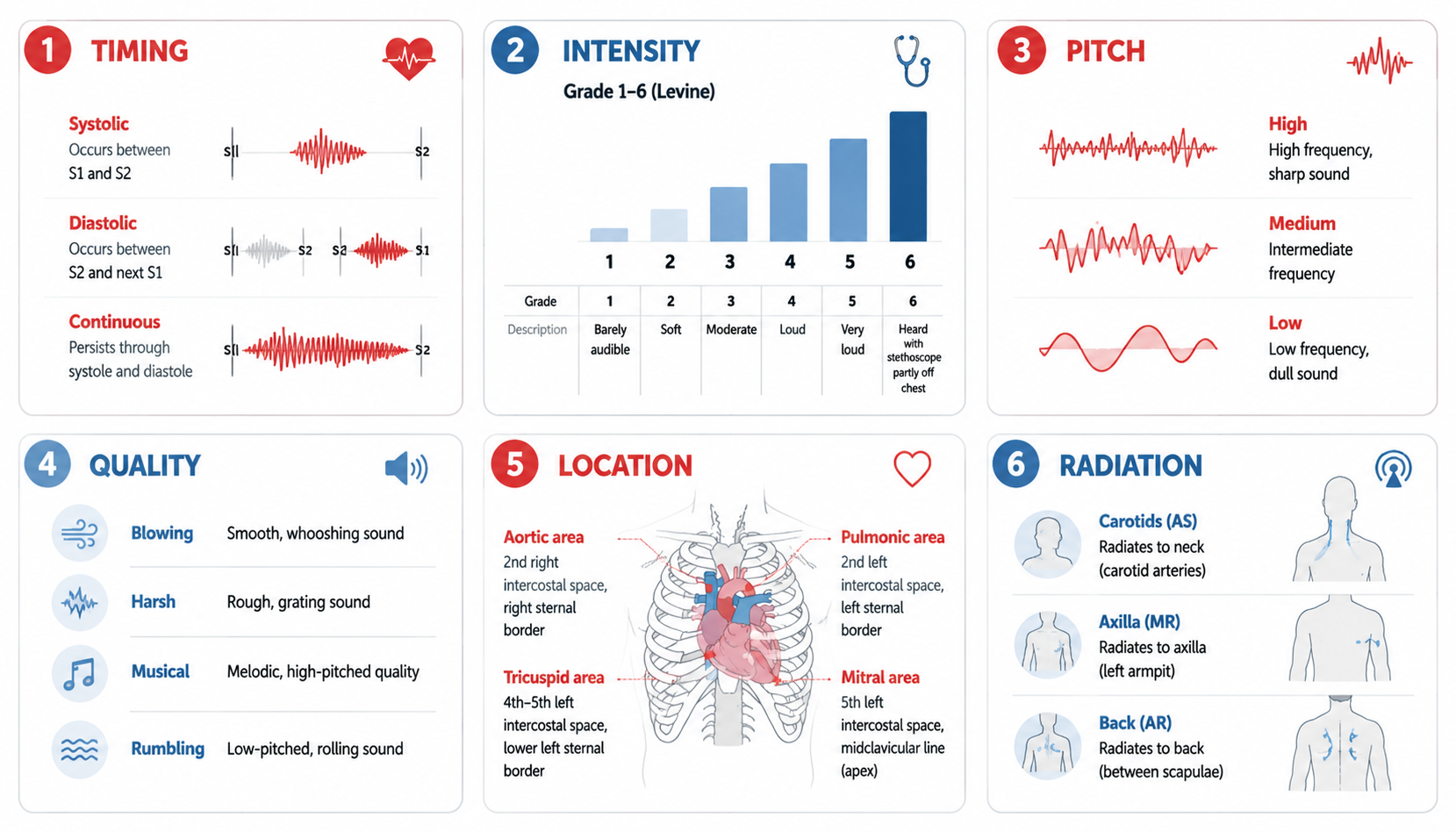
Core Concepts — Inspection & Palpation
Inspection
- Precordial bulge (long-standing cardiac enlargement, childhood disease)
- Apex beat: normally 5th ICS, 1–2 cm medial to MCL — displaced left/down = LV enlargement; invisible in obesity/emphysema
- Pulsations elsewhere (epigastric — RVH, COPD, AAA)
- General: scars (sternotomy = CABG/valve replacement), pacemaker, signs of heart failure (JVP, edema)
Palpation
- Apex beat: location, character (thrusting = LVH, tapping = MS), diffuse = dilatation
- Heave/lift: RVH (left parasternal), LVH (apex)
- Thrills (震颤): palpable murmurs — position + phase (systolic at apex = MR; at 2nd LICS = PS; diastolic = MS/AR); thrill = murmur grade ≥4
- Pericardial friction rub: palpable grating (pericarditis)
Core Concepts — Percussion
- Determines cardiac borders (relative dullness = real size; absolute dullness = no gas, over heart/liver)
- Normal borders: right 2–3 cm (II), 2–3 (III), 3–4 (IV) from midsternal line; left 2–3 (II), 3.5–4.5 (III), 5–6 (IV), 7–9 (V); midsternal to MCL 8–10 cm
- LV enlargement (aortic heart — boot shape): dullness extends left and downward — aortic insufficiency, hypertensive heart disease
- LA enlargement / pulmonary dilatation (mitral heart — pear shape): seen in mitral stenosis
- Pericardial effusion: both borders enlarge, changes with position
- Emphysema: cardiac dullness reduced
- Retromanubrial dullness ≥6 cm: aortic aneurysm, retrosternal goiter, thymic tumor, lymphoma
Core Concepts — Auscultation (听诊)
Valve areas — MUST KNOW
| Area | Location |
|---|---|
| Mitral (M) | 5th LICS, 1–2 cm medial to MCL (apex) |
| Pulmonary (P) | 2nd LICS just lateral to sternum (left) |
| Aortic (A) | 2nd RICS just lateral to sternum |
| Second aortic / Erb (E) | 3rd LICS lateral to sternum |
| Tricuspid (T) | 4th–5th ICS left sternal border |
- Routine order: M → P → A → E → T (counterclockwise; NOT equal to anatomic valve location)
- Stethoscope: diaphragm pressed tightly = high-pitched (valve closure, systolic events, regurgitant murmurs); bell touched lightly = low-pitched (gallops, AV stenosis murmurs)
Heart rate & rhythm
- Normal 60–100 bpm; <60 bradycardia, >100 tachycardia
- Sinus arrhythmia: faster on inspiration, slower on expiration — normal in young adults/children
- Premature beat: sudden extra systole + compensatory pause; bigeminy (every sinus beat + premature), trigeminy (every two); confirmed by ECG
- Atrial fibrillation — 3 irregularities: irregularly irregular rhythm, variable intensity of S1, pulse deficit (apical HR > radial pulse)
Heart sounds
| S1 | S2 | |
|---|---|---|
| Mechanism | Closure of AV valves (M+T) | Closure of semilunar valves (A+P) |
| Timing | Onset of ventricular systole (with apex beat/carotid pulse) | Onset of diastole |
| Pitch | Low | High |
| Intensity | Loud | Weak |
| Duration | Long (~0.1 s) | Short (~0.08 s) |
| Best heard | Apex | Base |
| Normal | A2 > P2 in old; P2 > A2 in children | — |
- Identify: S1 synchronous with carotid pulse/apex beat; diastole (S2→S1) longer than systole (S1→S2); move to base to identify S2 first
Extra heart sounds — Diastolic
| Sound | Timing | Cause |
|---|---|---|
| S3 (ventricular gallop, pathological) | Early diastole, low-pitched | Ventricular overload/failure — HF, acute MI, severe myocarditis, dilated CMP (physiologic S3 in children/young adults) |
| S4 (atrial gallop) | Late diastole, just before S1 | Atrial contraction against stiff/hypertrophic ventricle — LVH, long-standing HTN, AS, HCM |
| Summation gallop | S3+S4 fuse (fast HR) | HF, cardiomyopathies |
| Opening snap | Early diastole, sharp high-pitched | Mitral stenosis (flexible valve — suitable for commissurotomy) |
| Pericardial knock | Early diastole, left sternum | Constrictive pericarditis |
| Tumor plop | Diastole, positional | Left atrial myxoma |
Extra heart sounds — Systolic
| Sound | Cause |
|---|---|
| Early systolic click | Opening of deformed aortic/pulmonary valve (AS, pulmonary HTN); disappears when valve calcifies |
| Mid-late systolic click + late systolic murmur | Mitral valve prolapse (Barlow's syndrome) |
| Iatrogenic | Pacemaker sound, mechanical valve sound |
Cardiac murmurs — Mechanism
- Increased rate/velocity of flow (anemia, hyperthyroidism)
- Decreased valve diameter / constriction (MS, AS, PS)
- Valve insufficiency (MI, AI)
- Abnormal communication (ASD, VSD, PDA)
- Taut membrane vibration (vegetation, ruptured chordae)
- Sudden increase in vessel diameter (aneurysm)
Murmur description — 7 parameters
Location, Timing (phase), Quality, Intensity, Form, Radiation, Influencing factors (posture/exercise/respiration)
- Quality: blowing, rumbling, musical, machinery, harsh, jet-like, sigh-like, scratchy
- Intensity (Levine 1–6, systolic only): 1 = very faint; 2 = quiet but audible; 3 = moderately loud; 4 = loud + thrill; 5 = heard with stethoscope lightly on chest; 6 = heard with stethoscope off chest
- Grade 1–2 often physiologic; grade ≥3 rarely normal; diastolic murmurs are not graded
Murmur → Valve lesion map (HIGH YIELD)
| Valve lesion | Location | Phase | Quality/Radiation |
|---|---|---|---|
| AS | 2nd RICS (aortic area) | Systolic | Ejection crescendo-decrescendo, harsh → carotids; slow-rising pulse; S4 |
| AR | 3rd LICS (Erb) | Early diastolic | Blowing, decrescendo, leaning forward + expiration; water-hammer pulse, wide pulse pressure |
| MS | Apex | Mid-diastolic | Rumbling, low-pitched, bell; opening snap; tapping apex; LA enlargement (pear heart); S1 loud |
| MR | Apex | Pansystolic | Blowing, high-pitched → axilla; thrill possible; S3 |
| TR | 4th–5th LICS LSB | Pansystolic | ↑ with inspiration (Carvallo sign); JVP with prominent V wave |
| PS | 2nd LICS | Systolic | Ejection → radiates to back/neck |
| VSD | 3rd–4th LICS LSB | Pansystolic | Harsh, thrill |
| PDA | 2nd LICS | Continuous (machinery) | Through systole + diastole |
| Pericardial friction rub | LSB | Both phases | Grating, positional, breath-holding doesn't stop |
Auscultation key points
- S1 loud in MS; S1 soft in MR (valve not closing fully), AR
- Fixed split S2 = ASD; wide split = RBBB; paradoxical split = AS/HCM/LBBB; loud P2 = pulmonary hypertension
- Murmur changes: Valsalva ↓ most murmurs but ↑ HCM (obstruction); squat ↑ MR/VSD/AS, ↓ HCM
High-Yield Points
- 5 valve areas: M (apex, 5th LICS MCL), P (2nd LICS), A (2nd RICS), E/Erb (3rd LICS), T (4th–5th LICS LSB) — order M→P→A→E→T
- S1 = AV closure (with carotid/apex), low, long; S2 = semilunar closure, high, short
- Diastolic murmurs: MS = rumbling at apex + opening snap; AR = early diastolic blowing at Erb
- Systolic murmurs: AS = ESM at aortic area → carotids; MR = pansystolic at apex → axilla; VSD = pansystolic LSB; PS = ESM at pulmonary area; PDA = continuous machinery
- Thrill = murmur grade ≥4; palpable vibration
- A-fib: irregularly irregular + variable S1 + pulse deficit
- Gallops: S3 = ventricular failure/overload (HF, MI, DCM); S4 = stiff/hypertrophic ventricle (HTN, AS, HCM)
- Opening snap = MS (flexible valve); pericardial knock = constrictive pericarditis; tumor plop = LA myxoma
- Boot-shaped heart = aortic (AR, HTN); pear-shaped = mitral (MS); enlarged dullness = pericardial effusion
- Valve replacement scars: mid-sternotomy (CABG, AVR/MVR) — 70% of LMCHK CVS short cases are valve problems
LMCHK OSCE Practice
- CVS short case (8 min, Station 1): 70% valve problems, most commonly valve replacement — look for mid-sternotomy scar
- Sequence: general inspection (scars, clubbing, pallor, cyanosis, Marfan's) → hands (CRT, splinter hemorrhages, Osler's nodes, Janeway lesions) → pulse (rate, rhythm, volume, radio-femoral delay) → BP → face (xanthelasma, arcus senilis, malar flush of MS) → JVP (height, hepatojugular reflux) → precordium (apex beat, heaves, thrills) → auscultate M-P-A-E-T → auscultate carotids (AS radiation) → lungs bases (HF) → sacral/ankle edema → comment
- Common findings to name: aortic stenosis (slow-rising pulse, ESM → carotids), AR (water-hammer pulse, early diastolic), MS (loud S1, opening snap, diastolic rumble, tapping apex), MR (pansystolic → axilla)
- Comment format: "I can hear a pansystolic murmur, loudest at the apex, radiating to the axilla, consistent with mitral regurgitation"
- Heart murmurs video/audio: identify AS vs MS, AR vs MR (first-sound, timing, location)
Topic Summary
CVS exam: inspect (scars, apex), palpate (apex character, heaves, thrills), percuss (borders — boot = aortic, pear = mitral), auscultate (M-P-A-E-T). Know S1 vs S2 (timing/pitch/duration), the diastolic extra sounds (S3 = failure, S4 = stiff ventricle, opening snap = MS, knock = constrictive, plop = myxoma), systolic clicks (MVP), and the murmur map (AS/AR/MS/MR/TR/PS/VSD/PDA with location, phase, quality, radiation). A-fib = 3 irregularities + pulse deficit. This is the highest-yield chapter for both written exams and LMCHK OSCE short cases.