Preparatory Mindset
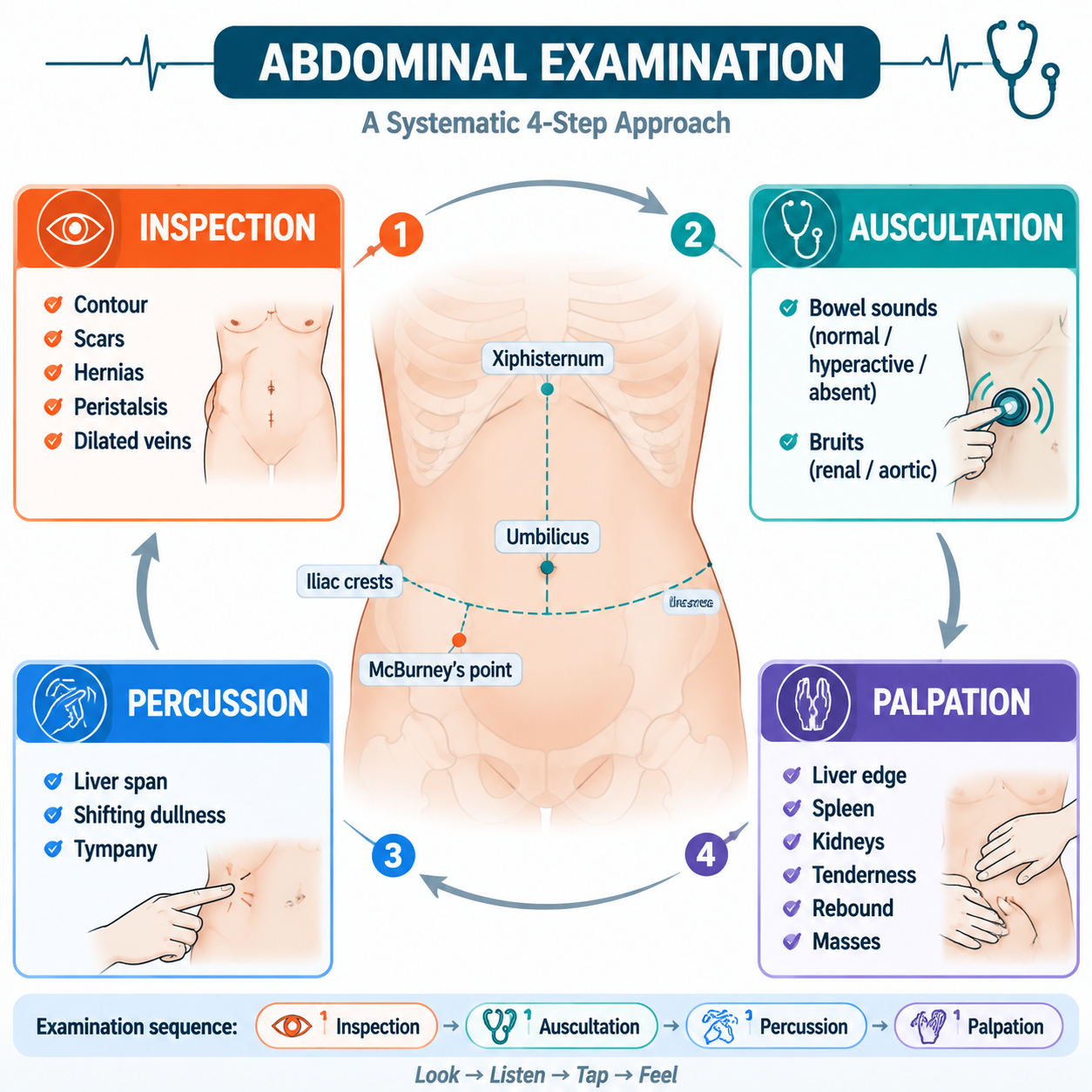
The abdominal examination follows a special order: Inspection → Auscultation → Percussion → Palpation (auscultation BEFORE percussion/palpation, unlike the chest). The patient lies flat, hips/knees flexed, examiner on the right. Master the 9-region/4-quadrant anatomy, the surface signs (Cullen's, Grey-Turner's, caput medusa), the liver/spleen/kidney palpation techniques, and the percussion findings (shifting dullness for ascites, Traube's space for splenomegaly). Always correlate with the common abdominal diseases and procedures (abdominocentesis).
Illustrations(图解速览)
Core Concepts — Anatomy & Preparation
Landmarks
Costal margin, xiphoid, umbilicus, anterior superior iliac spine, rectus abdominis lateral border, mid-abdominal line, inguinal ligament, pubic symphysis, costovertebral angle
Four quadrants / Nine regions
- Quadrants: RUQ (liver, gallbladder, hepatic flexure, right kidney), LUQ (spleen, stomach, splenic flexure, left kidney, pancreas tail), RLQ (cecum, appendix, right ovary/tube), LLQ (sigmoid, left ovary/tube)
- Nine regions (by costal arch + iliac spine lines × midclavicular lines): epigastric, left/right hypochondrium, umbilical, left/right lumbar, hypogastric, left/right iliac
Preparation
- Warm room, warm hands; supine, arms at sides, one pillow, hips/knees flexed; expose from pubis to costal margin (women: to below breasts)
- Stand on the patient's right side, face the patient
- Order: inspection → auscultation → percussion → palpation
Core Concepts — Inspection (视诊)
Abdominal contour
- Flatness (ideal level), fullness (above), bulge (obviously above), retraction/scaphoid (below — severe malnutrition, dehydration, cachexia, acute peritonitis)
- Whole bulge: ascites (frog abdomen — fluid sinks to flanks), pneumatosis (obstruction, ileus, perforation), large mass (ovarian cyst, teratoma); measure abdominal circumference at umbilicus
- Localized fullness: RUQ = liver/gallbladder; epigastric = stomach/pancreas/AAA (pulsatile); LLQ = sigmoid; suprapubic = bladder/uterus/ovary
- Masses by region: left subcostal = spleen/colon; RUQ = liver/gallbladder/pancreatic head; lumbar = kidney (cysts, tumor); RLQ = appendiceal abscess/cecal carcinoma; LLQ = diverticular abscess/sigmoid carcinoma; hypogastric = pelvic
- Abdominal wall vs intra-abdominal mass: ask patient to lift head off bed — if mass becomes more distinct = abdominal wall; if less = intra-abdominal
Respiratory movement
- Men & children: abdominal respiration; women: thoracic
- Peritonitis → absent or decreased abdominal wall movement (rigidity)
Abdominal veins & blood flow
- Caput medusa (dilated periumbilical veins radiating) = portal hypertension (umbilical vein collateral)
- IVC obstruction (Budd-Chiari): distended abdominal wall veins with cephalad flow
- Determine flow direction by emptying segment and refilling
Gastric/intestinal pattern & peristalsis
- Pyloric obstruction: peristalsis visible in epigastrium, left → right
- Intestinal obstruction: peristalsis around umbilicus, irregular
Other inspection findings
- Cullen's sign (periumbilical bluish) & Grey-Turner's sign (flank bluish) = retroperitoneal/intra-abdominal bleeding (severe pancreatitis, trauma, ruptured ectopic)
- Striae: silver (obesity/pregnancy), purple/bluish (Cushing's)
- Skin: rose spots (typhoid); pigmentation at belt line (Addison's)
- Hernias: umbilical (ascites), incisional (scar), femoral (female), inguinal (male)
- Hair distribution: male diamond (liver disease → female pattern); epigastric pulsation (thin, RVH/COPD, AAA)
Core Concepts — Auscultation (听诊)
Bowel sounds
- Normal: 4–5/min (gurgling)
- Increased/hyperactive: gastroenteritis, early obstruction, diarrhea (borborygmi)
- Decreased/absent: ileus, peritonitis, late obstruction
- Metallic tinkling: intestinal obstruction (fluid + air under tension)
Vascular bruits
- Abdominal aortic aneurysm: midline epigastric bruit + pulsatile mass
- Renal artery stenosis: systolic bruit in flanks/upper abdomen (hypertension)
- Hepatic bruit: hepatoma, AV malformation
- Venous hum (portal hypertension); friction rub over liver (perihepatitis/tumor); scratch test for liver edge
Core Concepts — Percussion (叩诊)
Liver
- Liver span: 9–12 cm in MCL (upper border at 5th ICS; lower border at costal margin)
- Dullness: enlarged liver (hepatitis, HF, tumor, cirrhosis early); upward displacement (ascites, pregnancy, tumor); loss of liver dullness + tympany = perforated viscus (free air under diaphragm)
- Floating liver ballottement in ascites
Spleen
- Traube's space (6th–9th ribs, left axillary to costal margin): normally tympanic
- Dullness in Traube's space = splenomegaly (or left pleural effusion, gastric mass)
- Splenic dullness: enlarge downward/medially; massive (leukemia, malaria, portal HTN, Gaucher's)
Kidney
- Costovertebral angle tenderness (pyelonephritis, stones)
Ascites — HIGH YIELD
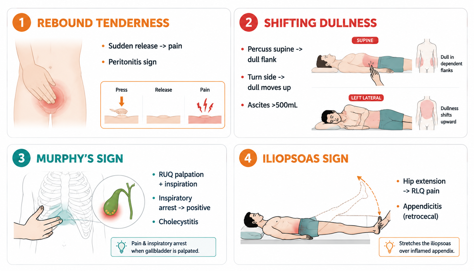
- Shifting dullness: dull flanks → tympanic center in supine; roll to side → dullness shifts to dependent side (ascites ≥1000–1500 ml)
- Fluid thrill/wave (波动感): assistant's hand edge on midline, tap flank → transmitted wave (large ascites)
- Succussion splash (振水音): shake patient → splash sound (gastric retention/obstruction, or soon after drinking)
- Bladder: suprapubic dullness = distended bladder (retention)
Core Concepts — Palpation (触诊)
Abdominal wall & tenderness
- Light palpation first (muscle tenseness, guarding); then deep
- Rebound tenderness (反跳痛): deep press 2–3 s, release quickly → pain intensifies = peritonitis (with rigidity)
- Specific points: McBurney (appendicitis), gallbladder point (cholecystitis), ureter points (stones)
- Muscle rigidity: generalized (perforation — board-like), localized (localized peritonitis)
Liver palpation
- Bimanual: left hand supports right lower ribs posteriorly; right hand flat on abdomen below costal margin; palpate during deep inspiration
- Describe: edge (sharp = normal; blunt/rounded = congestion), surface (smooth = hepatitis/HF; nodular = cirrhosis/tumor), consistency (firm = cirrhosis; hard = tumor), tenderness (HF, hepatitis, abscess — tender; tumor — non-tender), pulsatility (TR)
- Enlarged tender liver + JVD + hepatojugular reflux → right heart failure
- Hepatojugular reflux: press RUQ → JVP rises (RHF, constrictive pericarditis)
Spleen palpation
- Start from left lower quadrant (RIQ approach if massive), move up toward LUQ; palpate during inspiration
- Splenomegaly grading: mild (below costal margin 2–3 cm), moderate (to umbilicus), massive (>umbilicus — CML, myelofibrosis, malaria, kala-azar)
- Notch on medial border confirms spleen (vs renal mass)
Gallbladder & kidneys
- Courvoisier's sign: painless palpable enlarged gallbladder + jaundice = pancreatic head carcinoma (vs tender + jaundice = stones)
- Murphy's sign: press gallbladder point, ask deep inspiration → arrested breath (acute cholecystitis)
- Kidneys: bimanual palpation (19CM exam tested); ballotment; enlarged = polycystic disease, tumor, hydronephrosis
Abdominal masses
Describe: location, size, shape, surface, consistency, tenderness, pulsatility (AAA), mobility (with respiration — liver/spleen/kidney move; fixed = malignancy/adhesion), bruit (AAA, renal artery)
Fluid thrill & succussion splash
As above in percussion.
Core Concepts — Common Abdominal Diseases (快速识别)
- Acute cholecystitis: RUQ pain after fatty meal, Murphy's sign, fever; USS → gallstones
- Cholangitis: Charcot's triad (RUQ pain + jaundice + fever) — emergency, IV antibiotics + drainage
- Acute pancreatitis: epigastric pain → back, after alcohol/meal, vomiting, ↑amylase/lipase; Cullen's/Grey-Turner's (severe)
- Appendicitis: periumbilical → RLQ migration, McBurney tenderness, Rovsing sign, fever, leukocytosis
- Perforated ulcer: sudden severe epigastric, board-like rigidity, free air under diaphragm on erect CXR
- Intestinal obstruction: colicky pain + vomiting + distension + constipation; hyperactive then absent sounds; metallic tinkling
- AAA: pulsatile midline mass, tearing back pain, hypotension — surgical emergency
- Hepatic cirrhosis: ascites, caput medusa, spider angioma, splenomegaly, palmar erythema, gynecomastia
Core Concepts — Abdominocentesis (腹腔穿刺术)
- Indications: diagnostic (new-onset/unknown ascites), therapeutic (severe distension, SOB, oliguria), SBP diagnosis, intraperitoneal drugs/chemotherapy
- Contraindications: acute surgical abdomen (absolute); late pregnancy, ovarian cyst, hydatid, distended bladder, cellulitis, severe distended bowel, severe adhesions, restless patient (relative); bleeding risk if PT >21 s, INR >1.6, platelets <50,000
- Sites: ①outer 1/3 of line between umbilicus and ASIS (left/right); ②umbilicus horizontal line × anterior/mid-axillary line (lateral decubitus); ③1 cm above umbilicus-pubis midpoint, 1.5 cm lateral; ④ultrasound-guided; avoid scars
- Procedure: consent → empty bladder → supine, head elevated 20–30° → sterile prep (15 cm, 3×) → lidocaine → Z-tracking (prevent leak) → slow advance with aspiration → diagnostic 50 ml; therapeutic up to 3 L (cirrhosis) with albumin if >4–5 L
- Complications: fluid not flowing, feculent/bloody tap, hypotension after large-volume paracentesis (IV saline, albumin), infection, bowel perforation
- Ascitic fluid analysis: SAAG, protein, cell count, culture, cytology, ADA (TB)
High-Yield Points
- Abdomen: auscultate BEFORE percussion/palpation (unlike chest)
- Shifting dullness = ascites ≥1–1.5 L; fluid thrill = large ascites; succussion splash = gastric retention
- Rebound tenderness + rigidity = peritonitis
- Cullen's (periumbilical) & Grey-Turner's (flank) = retroperitoneal bleeding (pancreatitis, ectopic rupture)
- Caput medusa = portal hypertension; loss of liver dullness + tympany = perforated viscus
- Courvoisier's sign (painless palpable GB + jaundice) = pancreatic head cancer
- Murphy's sign = acute cholecystitis; McBurney tenderness = appendicitis; Charcot's triad = cholangitis
- Traube's space dullness = splenomegaly; massive spleen (below umbilicus) = CML/myelofibrosis/malaria
- Abdominocentesis: Z-tracking, empty bladder, 50 ml diagnostic, ≤3 L therapeutic (albumin if >4–5 L)
- Ascites: SAAG ≥1.1 = portal hypertension (cirrhosis, HF); <1.1 = exudative (TB, cancer, nephrotic)
LMCHK OSCE Practice
- Abdominal short case (8 min, Station 1): common cases — liver, kidney, haematological diseases; check for surgical scars (lobectomy/colectomy), stoma
- Sequence: consent + hand hygiene → general inspection (jaundice, anemia, cachexia, spider angioma) → inspect abdomen (contour, veins, scars, peristalsis) → auscultate (bowel sounds, bruits) → percuss (liver span, Traube's space, shifting dullness) → palpate (light → deep, liver, spleen, kidneys, masses) → check for ascites (shifting dullness, fluid thrill) → offer DRE/hernia examination → comment
- Liver: "liver edge palpable 3 cm below costal margin, firm, non-tender, smooth — consistent with hepatomegaly"
- Ascites: perform shifting dullness + fluid thrill; state you'd do USS + diagnostic paracentesis
- Acute abdomen OSCE: recognize peritonism, rebound tenderness; urgent surgical referral; don't give analgesia before surgical review (traditionally)
- Mention you would check: urine pregnancy test in women, blood glucose, ECG (MI presenting as epigastric pain)
Topic Summary
Abdominal exam order: inspection → auscultation → percussion → palpation. Inspection: contour (scaphoid/frog abdomen), veins (caput medusa), Cullen's/Grey-Turner's, peristalsis. Auscultation: bowel sounds (absent = ileus, tinkling = obstruction), bruits (AAA, renal artery). Percussion: liver span, Traube's (spleen), shifting dullness (ascites), loss of liver dullness (perforation). Palpation: rebound (peritonitis), liver (edge/surface/tenderness), spleen (notch, massive = CML), Murphy's (cholecystitis), Courvoisier's (pancreatic cancer). Procedures: abdominocentesis with Z-tracking and SAAG interpretation. Correlate with common diseases (appendicitis, pancreatitis, cholangitis, obstruction, AAA).