Preparatory Mindset
Bleeding disorders (出血性疾病) arise from three "hemostatic legs": vessel wall, platelets, and coagulation factors. Recognize the clinical patterns — mucocutaneous bleeding (petechiae/purpura) = vessel/platelet problem; deep hematoma/joint bleeding = coagulation disorder. Master the screening tests (BT, platelet count, PT, APTT, TT, FIB) and their interpretation, the DIC picture, and the bone marrow examination used to diagnose hematologic diseases (leukemia MICM, ITP, multiple myeloma, aplastic anemia).
Illustrations(图解速览)
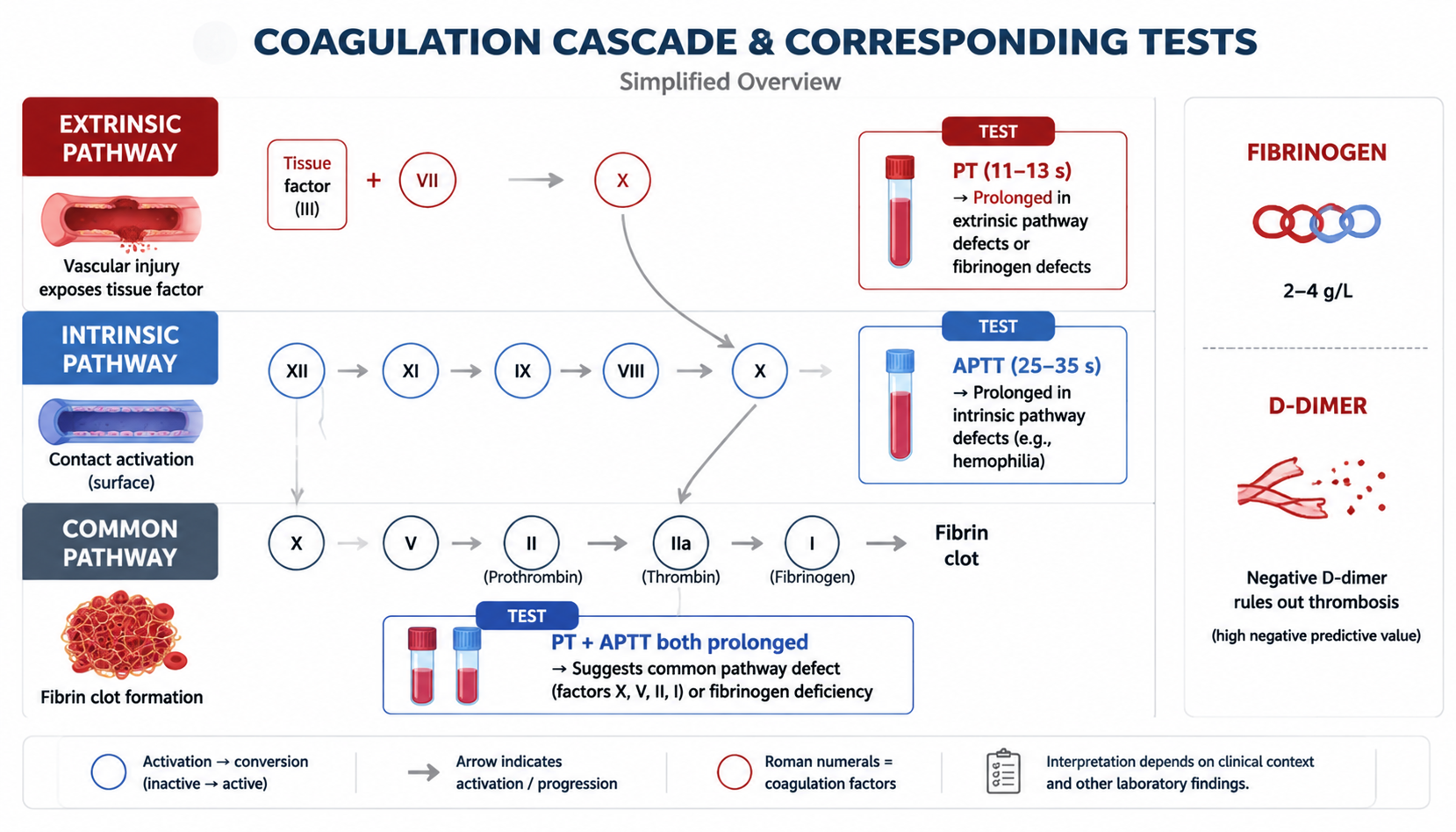
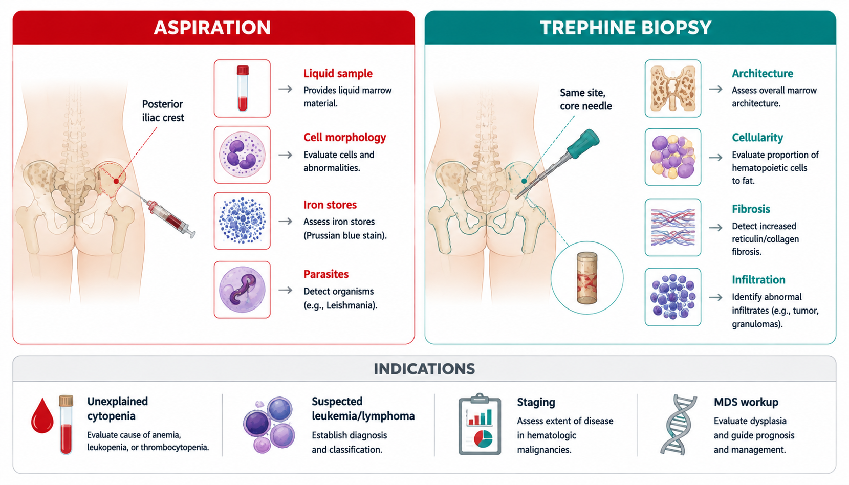
Core Concepts — Normal Hemostasis & Coagulation
Hemostasis mechanism (three legs)
- Vessel wall: reflex vasoconstriction; endothelial damage → release tissue factor (TF, extrinsic pathway), activate factor XII (intrinsic pathway), release vWF + endothelin
- Platelets: adhesion (vWF), aggregation, release → platelet plug
- Coagulation factors: cascade → fibrin clot (final: Fibrinogen → Fibrin via thrombin)
Coagulation pathways — MUST KNOW
| Pathway | Trigger | Screened by | Factors |
|---|
| Intrinsic (内源性) | Factor XII activation (contact with damaged endothelium) | APTT | XII, XI, IX, VIII (+common) |
| Extrinsic (外源性) | Tissue factor (TF) released by damaged tissue | PT | VII (+common) |
| Common (共同途径) | — | PT + APTT both | X, V, II (prothrombin), I (fibrinogen) |
- Intrinsic deficiency → APTT↑, PT normal (hemophilia A/B — factors VIII/IX)
- Extrinsic deficiency → PT↑, APTT normal (factor VII)
- Common pathway deficiency → PT↑ and APTT↑ (X, V, II, I)
- Vitamin K–dependent factors: II, VII, IX, X (+ proteins C, S) — deficiency in vitamin K deficiency, liver disease, warfarin → PT↑ (VII short half-life, first to drop)
- Liver disease: prolonged PT (all factors made in liver except VIII + vWF)
- Balance: anticoagulants (antithrombin III, protein C/S) limit clot to local area; overwhelming stimulation → DIC
Fibrinolysis
- Plasminogen → plasmin → dissolves fibrin clot
- Hyperfibrinolysis → bleeding (liver disease, DIC, tPA therapy)
Core Concepts — Mucocutaneous Hemorrhage (皮肤黏膜出血)
Clinical forms
| Form | Size/Feature |
|---|
| Petechiae (瘀点) | <2 mm |
| Purpura (紫癜) | 3–5 mm |
| Ecchymosis (瘀斑) | >5 mm |
| Hematoma (血肿) | Palpable deep collection |
| Epistaxis / gingival bleeding / oral bleeding | Mucosal |
Classification of bleeding diseases
- Vascular abnormality: congenital (hereditary hemorrhagic telangiectasia) + acquired — allergic purpura (Henoch-Schönlein purpura, HSP): type III hypersensitivity, children, after respiratory infection, symmetric lower-limb purpura + abdominal pain + arthralgia + renal involvement (hematuria)
- Platelet abnormality: count ↓ (ITP — immune thrombocytopenia, aplastic anemia, leukemia) or ↑ (primary thrombocythemia); quality (thrombasthenia/platelet dysfunction)
- Coagulation abnormality: hereditary — hemophilia A (VIII), B (IX); acquired — liver disease, vitamin K deficiency, anticoagulant excess
- Fibrinolytic abnormality
- Generalized: DIC
Clinical pattern — vessel/platelet vs coagulation — HIGH YIELD
| Feature | Vessel/platelet | Coagulation |
|---|
| Bleeding site | Skin, mucosa (petechiae, purpura, epistaxis, gum, menorrhagia) | Deep: hematoma, joint bleeding (hemarthrosis), muscle |
| Onset | Immediate after trauma | Delayed |
| Gender | Females (menorrhagia) | Hemophilia: males |
| Family history | Variable | Strong (X-linked) |
| Examples | ITP, HSP, thrombasthenia, scurvy | Hemophilia A/B, liver disease, warfarin, DIC |
Core Concepts — Screening Tests (筛选试验)
| Test | Normal | ↑/Abnormal means |
|---|
| Platelet count | 100–300 ×10⁹/L | <50: bleeding risk with trauma; <20: spontaneous bleeding |
| Bleeding time (BT) | 1–3 min (Ivy) | ↑ in platelet/vascular disorders (not coagulation) |
| PT (prothrombin time) | 11–13 s | ↑ extrinsic/common: VII, X, V, II, I deficiency; vitamin K deficiency; liver disease; warfarin; DIC |
| APTT (activated partial thromboplastin time) | 25–35 s | ↑ intrinsic/common: hemophilia A/B (VIII/IX), XI, XII; heparin; DIC; lupus anticoagulant |
| TT (thrombin time) | 16–18 s | ↑: fibrinogen deficiency/dysfibrinogenemia, heparin, DIC |
| Fibrinogen | 2–4 g/L | ↓: DIC, severe liver disease, fibrinogenolysis |
| D-dimer | <0.5 mg/L | ↑: DIC, thrombosis (DVT/PE), post-op, malignancy |
PT vs APTT interpretation — MUST KNOW
| Pattern | Disease |
|---|
| APTT↑ only | Hemophilia A/B (VIII, IX), XI deficiency, heparin, lupus anticoagulant |
| PT↑ only | Factor VII deficiency, early vitamin K deficiency / warfarin, mild liver disease |
| Both PT + APTT↑ | DIC, severe liver disease, vitamin K deficiency (advanced), common pathway deficiency (X, V, II, I) |
| Both normal | Platelet/vascular disorder, factor XIII deficiency (normal screen!), mild platelet dysfunction |
- Factor XIII deficiency: normal PT/APTT/BT but delayed bleeding after 24–48 h + poor wound healing — send urea solubility test
Core Concepts — DIC (弥散性血管内凝血)
- Widespread activation of coagulation → microthrombi (organ ischemia) + consumption of platelets/factors + secondary fibrinolysis → bleeding
- Causes: sepsis (gram-negative), obstetric emergencies (abruptio placentae, amniotic fluid embolism), malignancy (APL — promyelocytic leukemia), trauma, liver failure, snake bite
- Diagnosis — DIC screen: ↓platelets, PT↑, APTT↑, ↓fibrinogen, D-dimer↑↑, schistocytes on film, ↑FDP
- Clinical: bleeding (petechiae, oozing), thrombosis (DVT, organ infarct), shock, renal/hepatic failure
- Management: treat the cause (antibiotics, delivery, chemotherapy), supportive (FFP, cryoprecipitate, platelets), consider heparin only in selected thrombotic-predominant DIC
Core Concepts — Bone Marrow Examination (骨髓检查)
Purpose & indications
- Extract BM tissue/fluid for morphology, pathology, immunology, chromosome, genetics (MICM) — diagnose or evaluate hematologic disease
- Indications: unknown blood cell abnormalities; hepatosplenomegaly + lymphadenopathy; fever of unknown origin; monitor treatment/evaluate disease status; research
- Contraindications: hemophilia; DIC/severe bleeding; local infection; uncooperative pregnant women/infants
- Complications: bleeding, needle fracture, pain, infection
Puncture technique
- Sites: posterior/anterior iliac crest (common), sternum, tibia (children)
- Amount: 0.2 ml for smear (no anticoagulant); 2–5 ml for chromosome/immunophenotype/gene
- Smear stained Giemsa-Wright; examine low power (10×) + oil power (100×)
- Red marrow: flat bones (sternum, pelvis, ribs, vertebrae, scapulae, long-bone ends); yellow marrow: long-bone shaft (converts back in severe blood loss)
Bone marrow in common diseases
| Disease | Marrow finding |
|---|
| Acute leukemia | Blasts ≥20% (AML: Auer bodies; ALL: PAS+, terminal TdT) |
| CML | Hypercellular, myeloid hyperplasia, Philadelphia chromosome t(9;22) BCR-ABL |
| Chronic lymphocytic leukemia | Lymphocytosis |
| Multiple myeloma | Plasma cells >10%, rouleaux, lytic lesions, monoclonal Ig |
| ITP | Normal/increased megakaryocytes (destruction peripheral) |
| Aplastic anemia | Hypocellular, fatty marrow |
| Anemia | Erythroid hyperplasia (hemolytic/deficiency response) |
MICM classification of leukemia (modern)
- Morphology (FAB), Immunophenotype (flow cytometry: CD markers), Cytogenetics (chromosomes), Molecular genetics (genes)
- AML markers: CD13, CD33, MPO; ALL: CD19 (B), CD3 (T), TdT
- t(8;21) AML M2, t(15;17) APL (M3 — DIC risk, treat with ATRA), inv(16) M4Eo, t(9;22) CML/ALL
High-Yield Points
- Petechiae/purpura = vessel or platelet; hematoma/joint bleeding = coagulation
- APTT↑ only = hemophilia (VIII/IX) or heparin; PT↑ only = VII deficiency/warfarin early; both↑ = DIC, liver disease, vit K deficiency
- Vitamin K–dependent factors: II, VII, IX, X (+ protein C/S)
- Hemophilia A = factor VIII deficiency (X-linked, males, joint bleeding, APTT↑, PT normal)
- ITP: ↓platelets with normal/increased megakaryocytes (immune destruction); HSP: vascular purpura, children, type III hypersensitivity
- DIC: ↓platelets + PT↑ + APTT↑ + ↓fibrinogen + D-dimer↑ + schistocytes
- Bone marrow: 0.2 ml smear, iliac crest common; blasts ≥20% = acute leukemia
- MICM = morphology + immunophenotype + cytogenetics + molecular
- APL (M3): DIC risk — treat with ATRA (all-trans retinoic acid)
- Factor XIII deficiency: normal screening tests but delayed bleeding
LMCHK OSCE Practice
- Coagulation interpretation station: given PT/APTT/platelets → localize the defect (platelet/vessel vs intrinsic vs extrinsic vs common)
- Bleeding history: onset (childhood = congenital), site (mucocutaneous vs joint/muscle), drug exposure (aspirin, warfarin, heparin, NSAIDs), family history (X-linked hemophilia), liver disease/alcohol, recent infection (HSP, ITP), menorrhagia
- Exam: check for petechiae, purpura, ecchymosis, hematomas, hemarthrosis, gum bleeding; liver disease stigmata (spider angioma, splenomegaly)
- "This patient has mucosal bleeding with isolated APTT prolongation — I would suspect hemophilia or heparin effect and check mixing studies, factor VIII/IX levels"
- ITP: isolated thrombocytopenia, normal smear otherwise → rule out pseudothrombocytopenia (EDTA clumping), check anti-platelet antibodies
- Never miss DIC in: sepsis + bleeding + schistocytes → urgent workup and treat the cause
Topic Summary
Bleeding = vessel/platelet (mucocutaneous, immediate) vs coagulation (deep/joint, delayed). Coagulation cascade: intrinsic (APTT, VIII/IX/XI/XII), extrinsic (PT, VII), common (X/V/II/I); vitamin K factors II/VII/IX/X. Screening: BT/platelets, PT, APTT, TT, fibrinogen, D-dimer — interpret patterns to localize. DIC: consumption coagulopathy (↓plts, PT↑ APTT↑, ↓fibrinogen, D-dimer↑, schistocytes) — treat the cause. Bone marrow: indications/contraindications, iliac crest, Giemsa-Wright, blasts ≥20% = acute leukemia, MICM classification, marrow patterns of leukemia/myeloma/ITP/aplastic anemia.