Preparatory Mindset
The ECG is a non-invasive recording of cardiac electrical activity from 12 viewpoints. Approach it as a structured interpretation: rate → rhythm → axis → intervals → P wave → QRS → ST segment → T wave → QT. Normal conduction: SA node → atria → AV node → bundle of His → bundle branches → Purkinje fibers. Master the normal values (PR 0.12–0.20 s, QRS 0.06–0.12 s), heart rate calculation, axis determination, and the classic patterns of hypertrophy, ischemia/infarction, and arrhythmias.
Illustrations(图解速览)
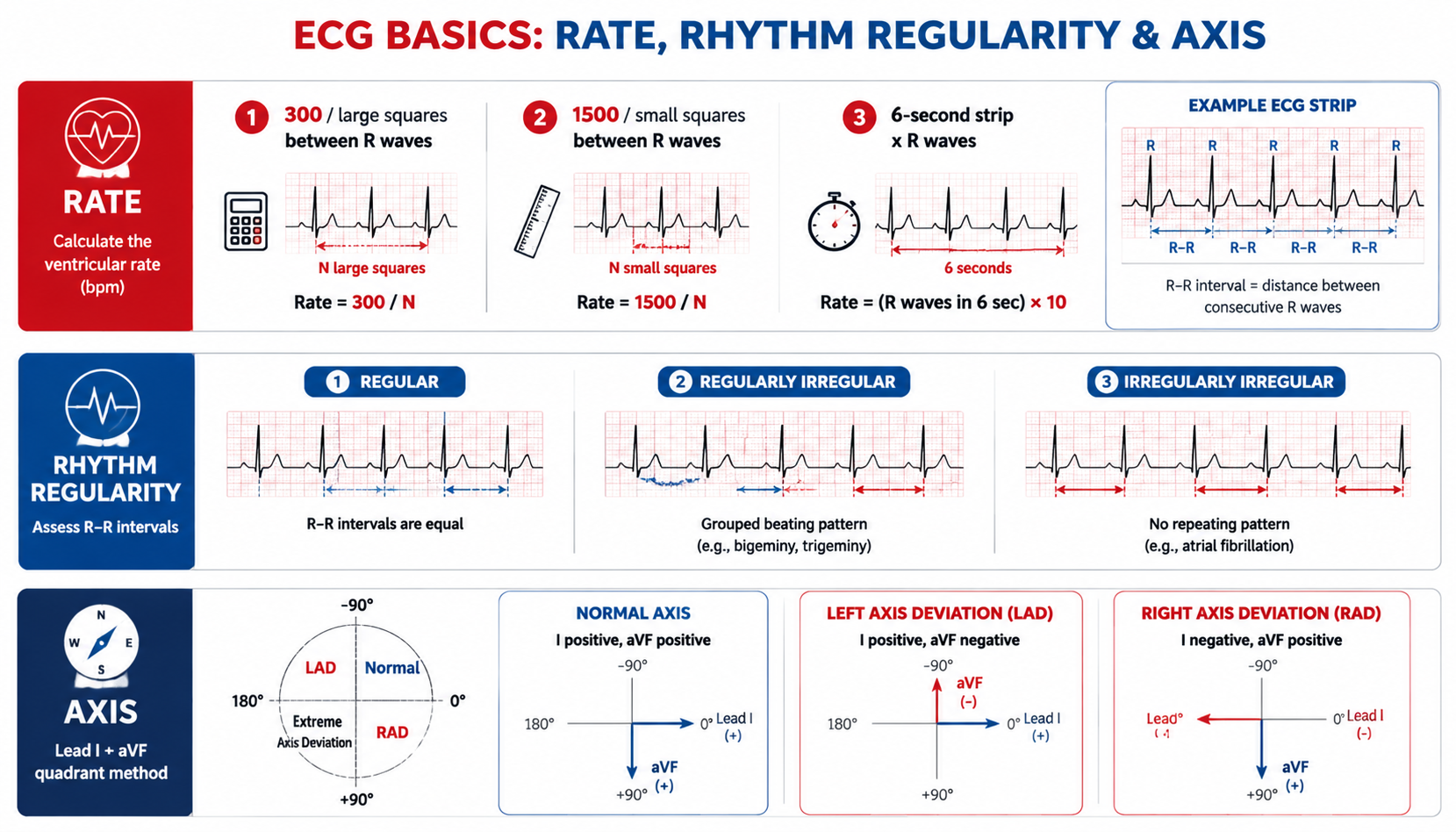
Core Concepts — Leads & Recording
12-lead system
| Group | Leads | Views |
|---|---|---|
| Standard (bipolar) | I, II, III | Frontal plane |
| Augmented | aVR, aVL, aVF | Frontal plane |
| Chest (precordial) | V1–V6 | Transverse plane |
- Limb leads (frontal): I, aVL → left lateral surface; II, III, aVF → inferior surface; aVR → right atrium
- Chest leads (transverse): V1, V2 → right ventricle; V3, V4 → septum + anterior LV; V5, V6 → anterior + lateral LV
Chest electrode positions — MUST KNOW
| Lead | Position |
|---|---|
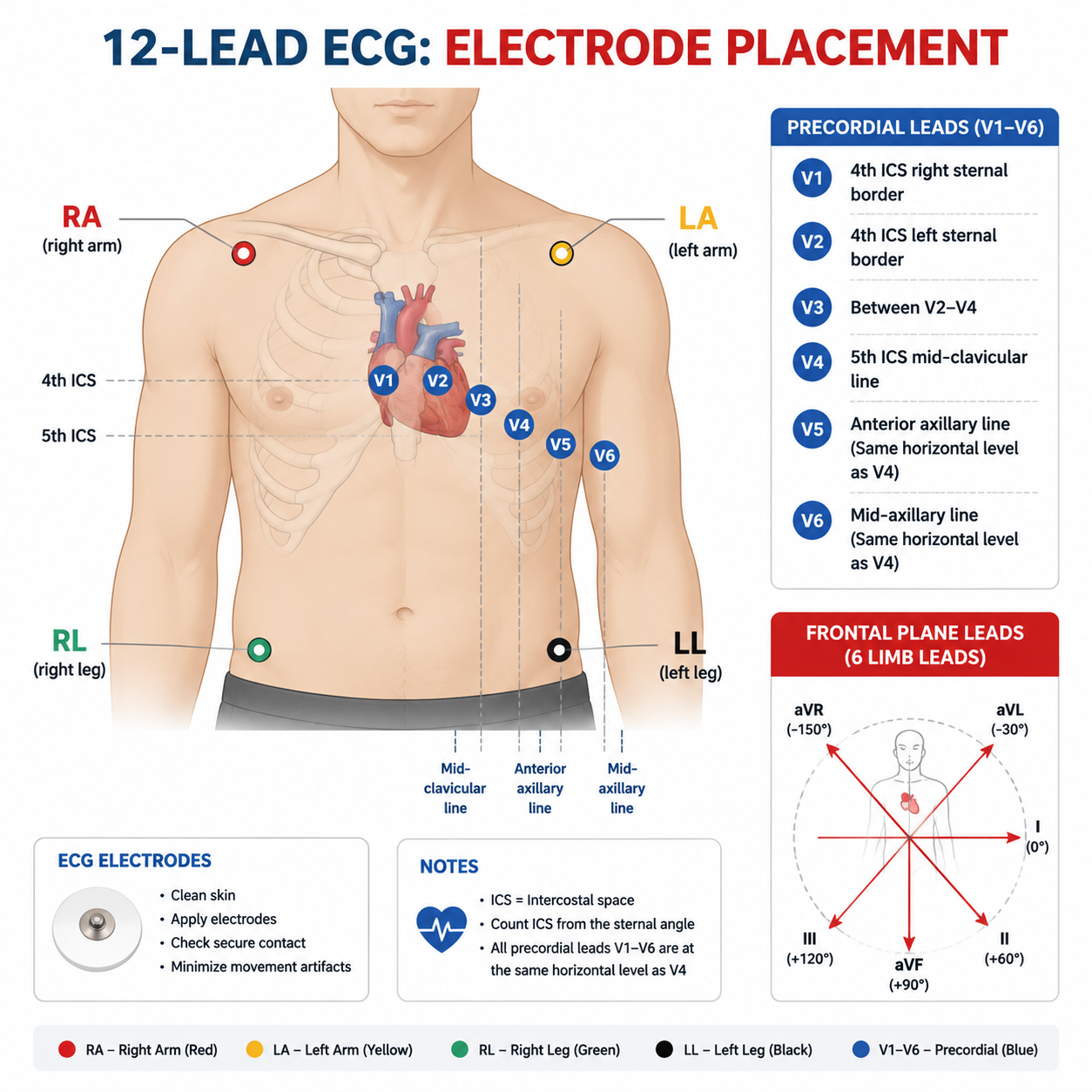
| V1 | Right 4th intercostal space (parasternal) |
| V2 | Left 4th intercostal space (parasternal) |
| V3 | Halfway between V2 and V4 |
| V4 | Left 5th ICS, midclavicular line |
| V5 | Horizontal to V4, anterior axillary line |
| V6 | Horizontal to V5, mid-axillary line |
Paper & calibration
- Speed 25 mm/s; voltage 10 mm/mV
- Small square (1 mm) = 0.04 s (horizontal), 0.1 mV (vertical); large square (5 mm) = 0.2 s, 0.5 mV
- Electrical impulse toward a lead → positive deflection; away → negative
Core Concepts — Normal ECG Interpretation
Normal sinus rhythm
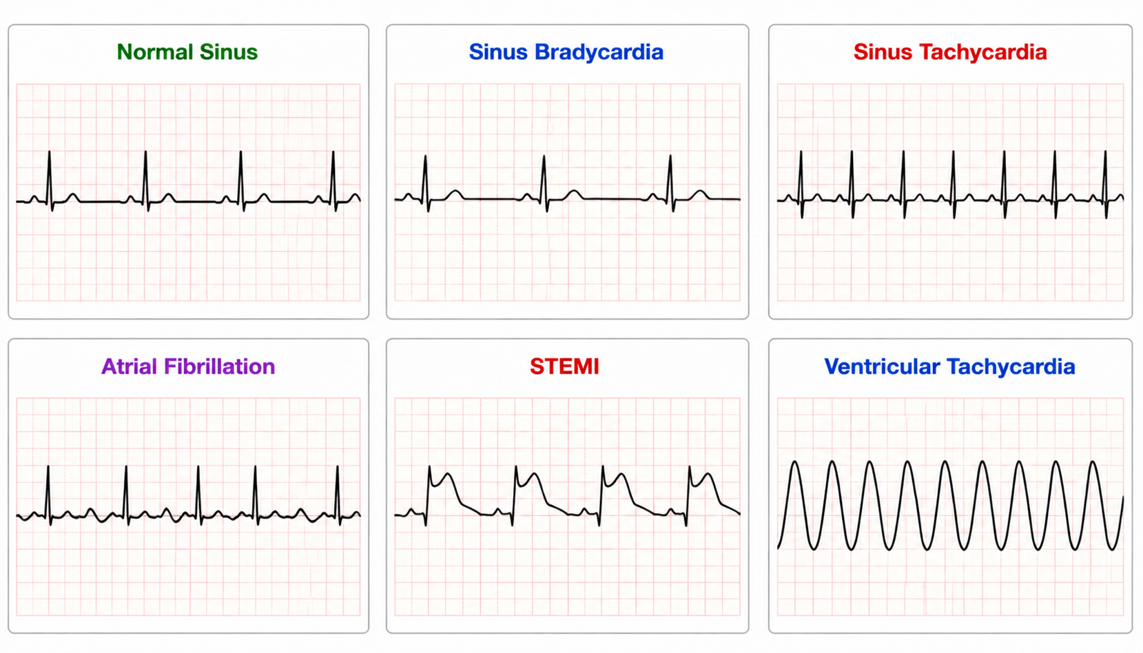
- Rate 60–100 bpm, regular, narrow QRS
- Each QRS preceded by P wave; P upright in lead II, down-going in aVR
- Conduction: SA node 60–100 bpm; AV junction 40–60; ventricle 20–40
Rate calculation
- Regular: HR = 60 ÷ (P-P or R-R interval in seconds) (e.g. R-R 0.7 s → 85 bpm)
- Alternate: 300 ÷ number of large squares between R waves
Waves & intervals — NORMAL VALUES
| Component | Represents | Normal |
|---|---|---|
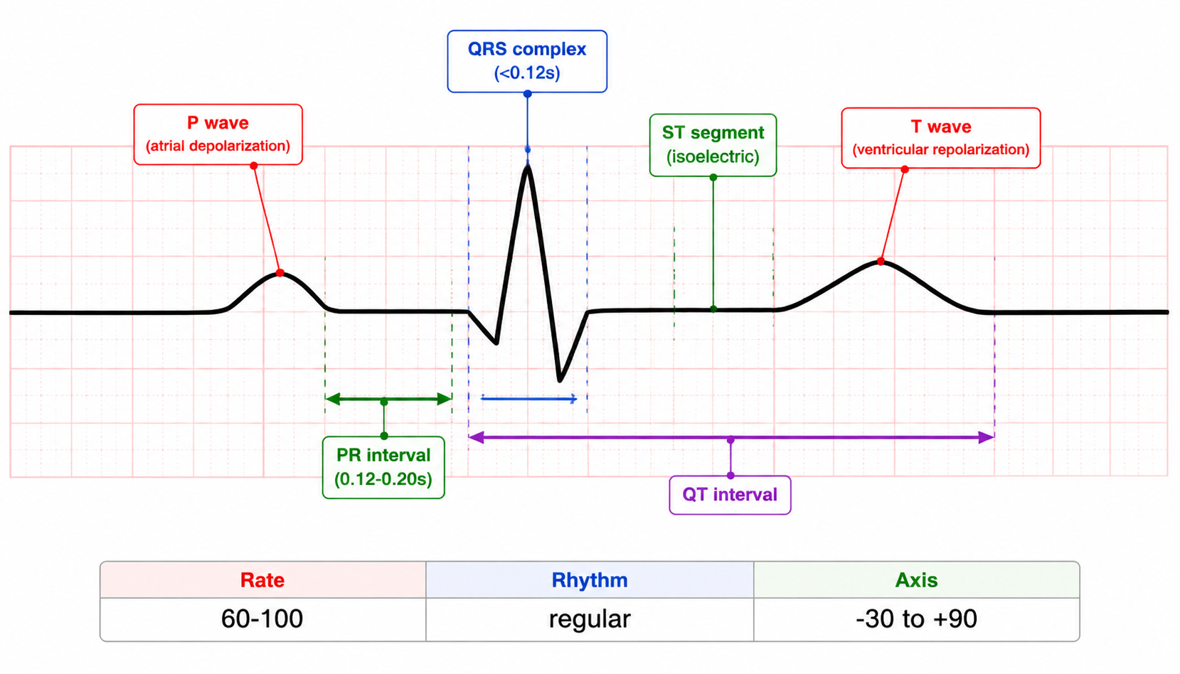
| P wave | Atrial depolarization | <0.12 s, amplitude <0.25 mV (2.5 mm); upright I, II, aVF; negative aVR |
| PR interval | Atrial → AV → ventricle | 0.12–0.20 s (>0.20 = 1st-degree AV block) |
| QRS | Ventricular depolarization | 0.06–0.12 s; Q = 1st negative, R = 1st positive, S = negative after R |
| ST segment | Isoelectric, depolarization → repolarization | Elevation/depression >0.1 mV (1 mm) = abnormal |
| T wave | Ventricular repolarization | Vector tracks QRS |
| QT interval | Electrical systole | <0.425 s corrected (QTc) |
Axis
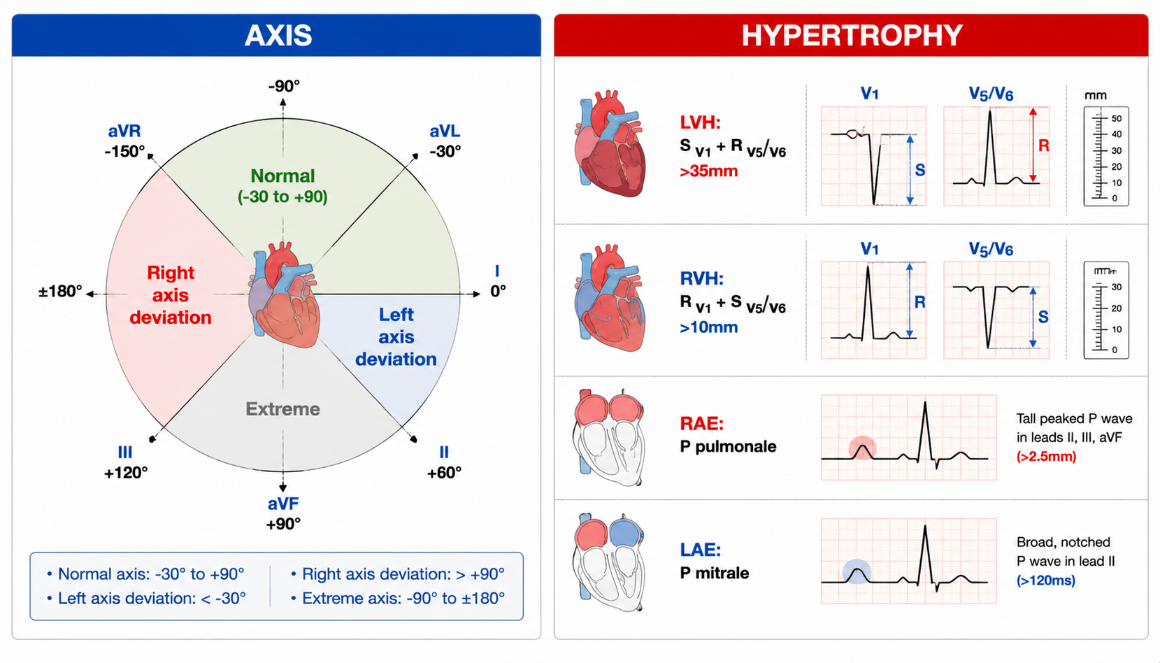
- Normal axis −30° to +90°
- Left axis deviation (LAD, −30° to −90°): left anterior hemiblock, LVH, inferior MI
- Right axis deviation (RAD, +90° to +180°): RVH, right bundle branch block, COPD, lateral MI, pulmonary embolism
Core Concepts — Chamber Enlargement & Hypertrophy
Atrial hypertrophy
- Left atrial (LAH): P wave broad, notched (duration >0.12 s) in I, II — "P mitrale" (mitral stenosis); biphasic P in V1 with terminal negative component (1)> (2)
- Right atrial (RAH): tall peaked P >0.25 mV in II, III, aVF — "P pulmonale" (COPD, pulmonary hypertension)
Ventricular hypertrophy
- LVH: tall R waves (R V5/V6 + S V1 >35 mm), LAD, LV strain (ST depression + T inversion in lateral leads)
- RVH: dominant R in V1 (R/S >1), RAD, right strain
Core Concepts — Arrhythmias
Bradycardia (<60 bpm)
- Sinus bradycardia (athletes, hypothyroidism, inferior MI)
- AV block: 1st degree (PR >0.20 s); 2nd degree (Mobitz I/Wenckebach — progressive PR prolongation then dropped QRS; Mobitz II — fixed PR, sudden dropped QRS); 3rd degree (complete dissociation, P and QRS independent)
- Sick sinus syndrome (elderly, syncope)
Tachycardia (>100 bpm)
- Sinus tachycardia (fever, pain, anemia, HF, hyperthyroidism)
- SVT: regular narrow complex, sudden onset — AVNRT most common; PSVT
- Atrial flutter: sawtooth flutter waves (II, III, aVF), atrial ~300, ventricular ~150 (2:1)
- Atrial fibrillation: irregularly irregular, no P waves, fibrillatory baseline
- VT: wide QRS >0.12 s, regular, rate 150–200 — medical emergency
- VF: chaotic, no output — cardiac arrest
- Torsades de pointes: polymorphic VT in long QT
Ischemia & Infarction — HIGH YIELD
- Ischemia: ST depression, T wave inversion (or hyperacute T in early injury)
- Injury (STEMI): ST elevation ≥1 mm (2 mm in V2-V3 men) in contiguous leads
- Infarction evolution: hyperacute T → ST elevation → T inversion → Q wave (pathologic: >1 mm wide or >1/3 R wave height) → ST returns
- Localization:
| Territory | Leads |
|---|---|
| Inferior | II, III, aVF (RCA) |
| Anteroseptal | V1–V4 (LAD) |
| Anterior | V3–V4 |
| Lateral | I, aVL, V5–V6 (LCx) |
| Posterior | V1–V3 reciprocal ST depression, tall R |
| RV | V4R |
High-Yield Points
- Normal values: PR 0.12–0.20 s; QRS 0.06–0.12 s; QT <0.425 s (QTc); rate 60–100
- V1–V6 positions (4th ICS right/left, V4 at 5th ICS MCL, V5-V6 axillary lines)
- NSR: P before each QRS, upright II, negative aVR
- HR = 60/R-R (s) or 300 ÷ large squares
- ST elevation = injury (STEMI) — act fast (primary PCI/fibrinolysis); reciprocal changes help localization
- LVH: R V5/V6 + S V1 >35 mm; RVH: R V1 > S V1
- P mitrale (LAH, MS) vs P pulmonale (RAH, COPD)
- 3rd-degree AV block: P-QRS dissociation — pacemaker
- A-fib: irregularly irregular, no P; VF = arrest; Torsades = long QT
- Pathologic Q wave = completed infarction
- Axis: LAD (−30 to −90) = LVH/hemiblock; RAD (+90 to +180) = RVH/COPD/PE
LMCHK OSCE Practice
- Non-interactive stations commonly include ECG interpretation: identify STEMI (ST elevation with reciprocal change), A-fib, complete heart block, SVT, VT, LVH, hyperkalemia (peaked T, wide QRS)
- Comment structure: rate → rhythm → axis → intervals → ischemia/infarction → conclusion + management
- "This ECG shows atrial fibrillation with a ventricular rate of ~110; the patient requires rate control and anticoagulation assessment (CHA2DS2-VASc)"
- STEMI: "ST elevation in leads II, III, aVF with reciprocal depression in I, aVL — inferior STEMI; activate the cath lab"
- Always correlate ECG with clinical picture and compare with previous ECG
Topic Summary
ECG = 12 leads (I-III, aVR-aVL-aVF, V1-V6) at 25 mm/s. Interpret systematically: rate (60/R-R), rhythm (P before QRS, upright II = sinus), axis (−30 to +90), intervals (PR ≤0.20, QRS ≤0.12, QTc <0.425), then P/QRS/ST/T/QT. Hypertrophy: P mitrale/P pulmonale, LVH/RVH voltage criteria. Arrhythmias: sinus/AV blocks/SVT/flutter/A-fib/VT/VF/Torsades. Ischemia-infarction: ST depression + T inversion (ischemia) vs ST elevation (injury/STEMI) with lead localization, then Q waves. ECG interpretation is a guaranteed OSCE non-interactive station.