Preparatory Mindset
This chapter covers the instrumental/procedural examinations of the respiratory and GI tracts: pulmonary function tests (PFT), bronchoscopy, and gastrointestinal endoscopy. Master the PFT pattern recognition (obstructive vs restrictive), the indications/contraindications of bronchoscopy, and the indications, preparation, and findings of upper/lower GI endoscopy. These are OSCE non-interactive station favorites — you must be able to comment on spirometry and name the endoscopic indications/complications.
Illustrations(图解速览)
Core Concepts — Pulmonary Function Testing (肺功能检查)
Components
- Lung volumes & capacities
- Spirometry: FEV1, FVC, FEV1/FVC, FEF25-75
- Flow-volume loop
- Bronchodilator response (reversibility)
- Diffusion capacity (DLCO)
- Bronchoprovocation testing (methacholine)
- Maximum respiratory pressures; cardiopulmonary exercise testing
Lung volumes — MUST KNOW
| Volume | Definition | Normal |
|---|---|---|
| Tidal volume (TV/VT) | Air per breath, quiet breathing | 6–8 ml/kg (~500 ml) |
| Inspiratory reserve volume (IRV) | Max inhaled from end-inspiratory tidal position | 1900–3300 ml |
| Expiratory reserve volume (ERV) | Max exhaled from resting end-expiratory position | 700–1000 ml |
| Residual volume (RV) | Air remaining after max exhalation — cannot be measured by spirometry | 20–25 ml/kg (1700–2100) |
Lung capacities
| Capacity | Composition | Normal |
|---|---|---|
| TLC | Sum of all volumes (IRV+TV+ERV+RV) | 4–6 L |
| VC | TLC − RV = IRV+TV+ERV | 60–70 ml/kg (3100–4800) |
| Inspiratory capacity (IC) | IRV + TV | 2400–3800 |
| Expiratory capacity (EC) | TV + ERV | — |
| FRC | RV + ERV (end-expiratory resting) — measured by helium dilution/nitrogen washout/plethysmography, not spirometry | 30–35 ml/kg (2300–3300) |
- FRC functions: oxygen store, buffer for steady PaO2, prevents atelectasis, minimizes work of breathing and V/Q mismatch (if closing capacity < FRC)
- FRC ↑ with: height, erect posture (30% more than supine), ↓lung recoil (emphysema); ↓ with: obesity, supine, muscle paralysis, restrictive disease, pregnancy, anesthesia; FRC doesn't change with age
Spirometry — the essentials
- Measures FEV1, FVC, FEV1/FVC, FEF25-75
- FEF25-75: mean expiratory flow in middle half of FVC — reflects small airway (<2 mm) function
- Highest of 3 reproducible measurements
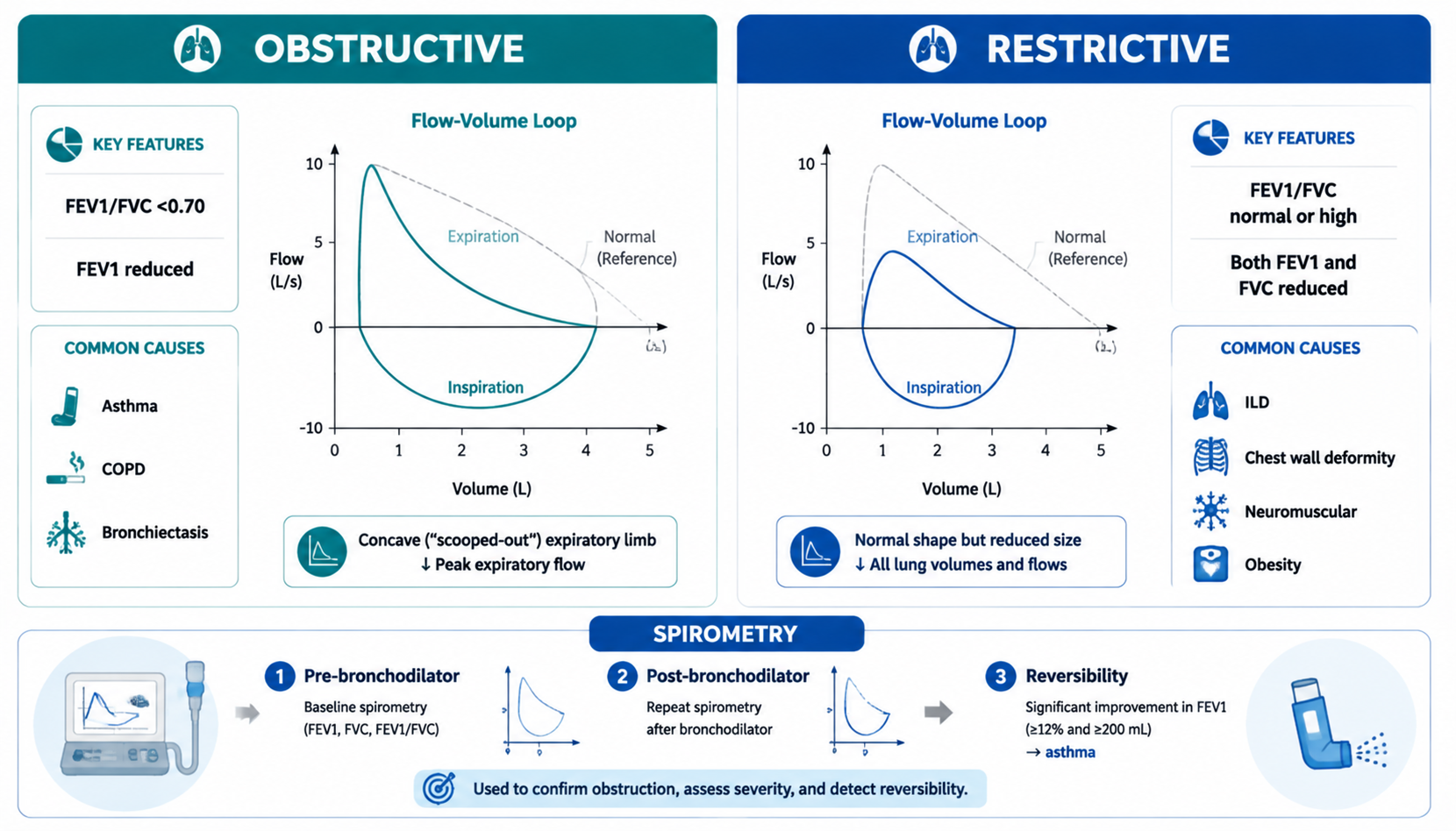
Interpretation — Obstructive vs Restrictive — HIGH YIELD
| Parameter | Obstructive (COPD, asthma, bronchiectasis, bronchiolitis) | Restrictive (fibrosis, obesity, kyphoscoliosis, effusion, neuromuscular) |
|---|---|---|
| FEV1 | ↓↓ | ↓ |
| FVC | ↓ (maybe) | ↓↓ |
| FEV1/FVC | <70% (decreased) | Normal or increased |
| TLC | ↑ (emphysema) or normal | ↓ |
| RV | ↑ | ↓ |
| DLCO | ↓ (emphysema) / normal (asthma) | ↓ (fibrosis) |
| Causes | Asthma (reversible), COPD (chronic bronchitis/emphysema), bronchiectasis | Interstitial lung disease/fibrosis, chest wall, neuromuscular, pleural, ascites |
- FEV1/FVC <70% = obstructive; FEV1/FVC normal/high + ↓FVC = restrictive
- FEV1 grades severity in COPD (GOLD: FEV1 ≥80, 50–79, 30–49, <30)
- Bronchodilator response (reversibility): FEV1 ↑ ≥12% AND ≥200 ml = significant (asthma)
- DLCO: ↓ in emphysema and interstitial fibrosis; normal in asthma/COPD-chronic bronchitis
- Bronchoprovocation (methacholine): airway hyperresponsiveness — asthma diagnosis when spirometry normal
VC decreases with
- Muscle weakness (drugs, neuromuscular disease, cerebral tumors), pulmonary disease (pneumonia, bronchitis, asthma, fibrosis, emphysema, edema), space-occupying (tumor, effusion, kyphoscoliosis), abdominal (tumor, ascites), opioids, abdominal pain/splinting, posture
Core Concepts — Bronchoscopy (支气管镜)
Definition & types
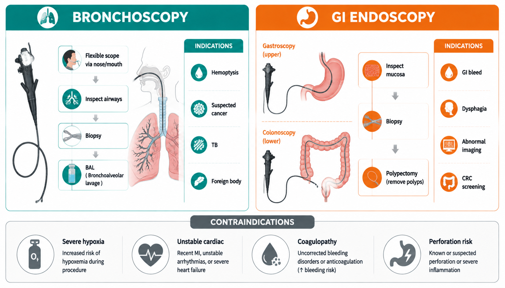
- Insertion of an endoscope into the bronchi to inspect the airway, remove objects, collect samples, place devices
- Rigid bronchoscope: straight hollow metal tube, general anesthesia, larger airway only — removal of foreign material, control of bleeding, stents; useful when bleeding obscures view
- Flexible (fiberoptic): bends to reach smaller airways, local anesthesia, more common — suction, biopsy, medicine delivery, breathing tube placement
- History: Gustav Killian 1897 (first), Shigeto Ikeda (flexible fiberscope)
Indications
- Lesions of unknown etiology on CXR; evaluate atelectasis or infiltrates
- Investigate hemoptysis, unexplained cough, wheeze or stridor
- Remove abnormal tissue or foreign material
- Retrieve foreign body
- Obtain lower respiratory secretions (washings, BAL), biopsy
- Determine location/extent of inhalation or aspiration injury
Contraindications
- Absolute: no informed consent (unless emergency + incompetent); no experienced bronchoscopist; inability to oxygenate
- Relative: lack of cooperation; recent MI/unstable angina; partial tracheal obstruction; moderate-severe hypoxemia; hypercapnia; pulmonary hypertension; uncorrectable coagulopathy; unstable hemodynamics/arrhythmias
Procedure essentials
- Premedication: benzodiazepines (midazolam) for anxiety; atropine to dry airway, promote anesthetic deposition, reduce vagal responses (bradycardia); opioids (morphine/fentanyl); bronchodilator before
- Airway preparation: lidocaine — gel to nasopharynx, atomizer to nose, mouthwash to oropharynx, nebulizer/scope to lower airways (prevent bleeding, cough, gagging, pain)
- Monitoring: continuous — oxygenation, vitals until pre-sedation level
- Complications: hypoxemia, bleeding (biopsy), laryngospasm, pneumothorax, arrhythmia, infection
Core Concepts — Gastrointestinal Endoscopy (内镜检查)
Upper GI endoscopy (EGD, gastroscopy)
- Indications: dyspepsia/ulcer symptoms (alarm features: age >50, weight loss, anemia, dysphagia, GI bleeding, persistent vomiting), upper GI bleeding (urgent), suspected tumor, stricture dilation, variceal banding, polypectomy, Barrett's surveillance, foreign body removal, biopsy (H. pylori, celiac)
- Preparation: NPO 8 h (fasting 6–8 h); stop anticoagulants when feasible; sedation (midazolam) optional
- Findings: esophagitis, Barrett's, varices, gastritis, gastric/duodenal ulcer (benign vs malignant margins), gastric cancer, polyp, angiodysplasia, Mallory-Weiss tear
- Complications: perforation (most serious), bleeding (post-polypectomy/biopsy), aspiration, sedation-related, infection
Lower GI endoscopy (colonoscopy)
- Indications: colorectal cancer screening (age 45–50), hematochezia/melena of lower origin, chronic diarrhea/IBD assessment (UC/Crohn's), polyp surveillance, change in bowel habit, iron-deficiency anemia, abnormal imaging
- Preparation: bowel prep (polyethylene glycol/clear fluid diet + laxatives day before) — cleanliness determines diagnostic yield; sedation; air/CO2 insufflation
- Findings: polyps (adenoma — premalignant; hyperplastic), diverticulosis, colorectal cancer, UC/Crohn's (continuous vs skip lesions, pseudopolyps vs cobblestone), angiodysplasia, hemorrhoids
- Complications: perforation, post-polypectomy bleeding (delayed up to 2 weeks), sedation complications, infection
- Sigmoidoscopy: distal colon/rectum — screening alternative
Other endoscopic modalities
- ERCP: endoscopic retrograde cholangiopancreatography — stones, strictures, stent, biliary decompression (complication: post-ERCP pancreatitis)
- EUS (endoscopic ultrasound): staging GI tumors, pancreatic lesions, FNA
- Capsule endoscopy: small bowel bleeding/lesions
- Polypectomy: complete removal, histology (adenomatous vs serrated), surveillance interval by size/histology
- Endoscopic hemostasis: injection (adrenaline), thermal, clips, banding (varices), argon plasma coagulation
Endoscopic red flags / findings to state
- Ulcer: benign (smooth, regular margin) vs malignant (irregular, raised, rolled edges, surrounding infiltration) — always biopsy
- Varices: dilated submucosal veins — band ligation, beta-blocker prophylaxis
- Barrett's esophagus: salmon-pink mucosa above GEJ — surveillance (dysplasia → ablation)
- Adenoma: premalignant — polypectomy + surveillance
- Colorectal cancer: irregular mass, stricture, bleeding — biopsy, staging CT
High-Yield Points
- Obstructive: FEV1/FVC <70% (↓); Restrictive: FEV1/FVC normal/↑ with ↓FVC and ↓TLC
- RV and FRC cannot be measured by spirometry (helium dilution/nitrogen washout/plethysmography)
- Reversibility: FEV1 ↑ ≥12% + ≥200 ml = asthma
- DLCO ↓ in emphysema + fibrosis; normal in asthma/chronic bronchitis
- FEF25-75 = small airway function
- Bronchoscopy: hemoptysis workup, biopsy, foreign body; rigid for foreign body/bleeding; flexible more common, local anesthesia
- Bronchoscopy contraindications: no consent, inability to oxygenate, recent MI, uncorrectable coagulopathy, severe hypoxemia/hypercapnia
- EGD for upper GI bleeding (urgent), colonoscopy for screening (45–50), ERCP complication = pancreatitis
- Colonoscopy prep quality determines yield; post-polypectomy bleeding can be delayed
- Always biopsy ulcers/masses; Barrett's needs surveillance
LMCHK OSCE Practice
- Non-interactive stations: comment on spirometry (obstructive vs restrictive, severity by FEV1), CXR of pneumothorax, CT of stroke/SAH, clinical photos (gangrene), X-rays (Colles', hip, ankle)
- "This spirometry shows reduced FEV1/FVC ratio of 58% with FEV1 60% predicted — obstructive pattern, consistent with COPD; I would perform bronchodilator testing to assess reversibility"
- Restrictive: "reduced FVC with normal FEV1/FVC and reduced TLC — restrictive pattern; consider interstitial lung disease"
- Endoscopy cases: CRC screening (polypectomy), achalasia (EGD finding), GI bleeding (urgent EGD ± variceal banding)
- Colon cancer: change in bowel habit + rectal bleeding + anemia in elderly → colonoscopy urgently
- Hemoptysis: CXR → CT → bronchoscopy; always exclude TB and tumor
Topic Summary
PFT: volumes (RV/FRC not spirometry), spirometry (FEV1/FVC), bronchodilator response, DLCO, bronchoprovocation — classify obstructive (FEV1/FVC <70%) vs restrictive (FVC↓, ratio normal/↑, TLC↓). Bronchoscopy: indications (hemoptysis, biopsy, foreign body), types (rigid vs flexible), contraindications, premedication (atropine, lidocaine). GI endoscopy: EGD (upper GI bleeding, ulcers, varices), colonoscopy (CRC screening, polyps, IBD), ERCP (pancreatitis risk), EUS (staging); complications (perforation, bleeding). Interpret patterns and name indications/complications fluently — classic OSCE non-interactive content.