Preparatory Mindset
Head & neck examination is a structured regional exam covering the skull, eyes, ears, nose, mouth, and neck (lymph nodes, thyroid, trachea, vessels) (19CM exam tested). It is heavily tested in OSCEs (thyroid, parotid, lymph nodes, fundoscopy, cranial nerves). Master the systematic sequence and the key signs: pupillary reflexes, papilledema, thyroid bruit, JVP, tracheal deviation, and lymph node characterization.
Illustrations(图解速览)
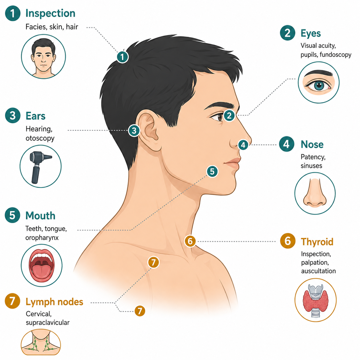
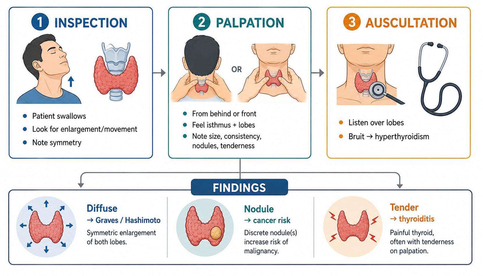
Core Concepts — Head
Skull & Scalp
- Size: normal, large (hydrocephalus — sunset eyes), small (microcephaly)
- Shape: acromegaly (enlarged, prominent brow), Paget's (enlarged, hat size ↑), rickets (frontal bossing), syphilis (saddle nose)
- Tenderness (temporal arteritis), masses, scars
- Scalp: seborrheic dermatitis, pediculosis, alopecia
Eyes — Detailed
| Structure | Normal | Abnormal findings |
|---|---|---|
| Eyelids | Symmetric, no swelling | Ptosis (CN III, Horner's, myasthenia), lid lag (hyperthyroidism), xanthelasma, edema (renal) |
| Conjunctiva | Pink, moist | Pallor (anemia), injection (conjunctivitis), petechiae (endocarditis), jaundice (sclera) |
| Sclera | White | Jaundice (bilirubin — liver/biliary), blue sclera (osteogenesis imperfecta) |
| Cornea | Clear | Arcus senilis (dyslipidemia), Kayser-Fleischer ring (Wilson's) |
| Pupils | Equal, round, reactive (PERRLA) | Anisocoria, fixed/dilated (CN III, herniation), constricted (pontine, opiates), RAPD (Marcus Gunn — optic nerve lesion) |
| Fundus | Disc, vessels, macula | Papilledema (↑ICP), Roth spots (endocarditis), AV nipping + hemorrhages (HTN), cotton wool spots + neovascularization (DM) |
| Extraocular | Full movement | Nystagmus, CN III/IV/VI palsy (diplopia), proptosis (thyroid, orbital tumor) |
- Pupillary light reflex: direct + consensual; afferent CN II, efferent CN III
- Visual field testing (confrontation): hemianopia patterns localize lesion (see neuro chapter)
- Fundoscopy technique: ophthalmoscope, red reflex first, follow vessels to disc, examine four quadrants
Ears, Nose, Mouth
- Ears: auricle (deformity, gouty tophi), external canal discharge, mastoid tenderness, hearing (whisper/Rinne/Weber)
- Nose: shape, nasal flaring (distress), discharge, septal deviation, sinus tenderness (frontal/maxillary)
- Mouth: lips (cyanosis, pallor, herpes, angular stomatitis), mucosa (petechiae, Koplik spots, leukoplakia), gums (bleeding — leukemia/periodontitis, lead line), tongue (cyanosis, atrophy — B12/iron, deviation — CN XII), teeth (caries, Hutchinson's — congenital syphilis)
- Tonsils/pharynx: injection, exudate, enlargement (grading: I within pillars, II beyond, III midline), palatal movement (CN IX/X), gag reflex
- Parotid gland: enlargement (mumps, stone, tumor) — examine opening of duct
Core Concepts — Neck
Sequence
Inspection → Palpation (lymph nodes, thyroid, trachea, vessels) → Auscultation (carotid bruits, thyroid bruit) → Assess JVP, neck stiffness, movements.
Lymph Nodes
- Cervical groups: submental, submandibular, anterior/posterior cervical, jugulodigastric, supraclavicular, occipital, post-auricular
- Characteristics: size, tenderness, consistency (hard = malignancy, rubbery = lymphoma, fluctuant = abscess/TB), mobility, matting
- Tender + small + mobile → infection; hard + fixed + non-tender → metastasis; generalized → lymphoma, leukemia, TB, HIV, syphilis, SLE
- "Describe a mass" in OSCE: site, size, shape, surface, consistency, tenderness, mobility, temperature, pulsatility, cough impulse (hernia), transillumination (cyst), bruit (vascular)
Thyroid Gland
- Inspection: midline neck mass, moves with swallowing
- Palpation: from behind, isthmus + both lobes; note size (grade I: palpable not visible; II: visible on swallowing; III: visible at rest), consistency (soft = goiter, firm = thyroiditis, hard/fixed = carcinoma), nodules, tenderness, thrill (bruit in Graves')
- Auscultation: systolic bruit in hyperthyroidism (Graves')
- Associated signs: exophthalmos, lid lag (Graves'); hoarseness (recurrent laryngeal nerve — carcinoma); retrosternal extension (obstruction)
- Thyroid function: TSH, free T3/T4; ultrasound; FNA for nodules
Trachea & Vessels
- Tracheal position: index finger in suprasternal notch, space equal both sides; deviation away from lesion = mass effect (pneumothorax, effusion, tumor); deviation toward = volume loss (atelectasis, fibrosis, post-pneumonectomy)
- Jugular venous pulse (JVP): internal jugular vein — assess at 45°; normal ≤4 cm above sternal angle; elevated JVP = right heart failure, pulmonary hypertension, SVC obstruction, constrictive pericarditis
- Hepatojugular reflux: press right upper quadrant → JVP rises (right heart failure, constrictive pericarditis)
- Carotid pulse: character (water-hammer = AR; slow-rising = AS; weak = shock), bruits (stenosis)
- Carotid vs JVP: carotid palpable, single waveform; JVP not palpable, double waveform, falls with inspiration, rises with abdominal pressure
Neck Movements & Meningeal Signs
- Range of motion: stiffness (meningitis, cervical spondylosis, RA)
- Kernig sign: flex hip 90°, extend knee → pain/resistance (meningitis)
- Brudzinski sign: flex neck → hips/knees flex (meningitis)
High-Yield Points
- Papilledema = ↑ICP (fundoscopy non-negotiable in headache/comatose patient before LP!)
- RAPD (Marcus Gunn) = afferent optic nerve lesion — swinging flashlight test
- Ptosis: CN III palsy (down/out eye, dilated pupil), Horner's (miosis + anhidrosis), myasthenia (fatigable, bilateral)
- Arcus senilis <50 years → dyslipidemia; Kayser-Fleischer ring → Wilson's
- Thyroid: moves with swallowing; bruit = Graves'; hard fixed = cancer; retrosternal = obstruction
- Tracheal deviation: away = mass, toward = collapse
- JVP ↑ = RHF/PHTN/constrictive pericarditis/SVC syndrome; hepatojugular reflux positive
- Lymph node: supraclavicular → malignancy; generalized → lymphoma/leukemia/TB/HIV/SLE
- Kernig + Brudzinski = meningeal irritation
- Parotid exam & "describe a mass" template are classic OSCE stations
LMCHK OSCE Practice
- Head & neck interactive stations (LMCHK surgery): thyroid exam, parotid exam, describe a mass, differential of lymph nodes, parotid gland swelling
- Thyroid exam sequence: consent + hand hygiene → inspection (midline mass, moves on swallowing, scars) → palpation from behind (isthmus, lobes, consistency, nodules, thrill) → auscultation (bruit) → check for retrosternal extension and tracheal deviation → cranial nerves (recurrent laryngeal — voice)
- Parotid: inspection (pre-auricular swelling, angle of jaw), palpation (bimanual — inside mouth + outside), examine duct opening (Stensen's), CN VII function (facial nerve runs through parotid!)
- Describe a lump: site, size, shape, surface, consistency, tenderness, mobility, fixity, temperature, pulsatility, transillumination, cough impulse, auscultation (bruit), overlying skin
- Eye exam: visual acuity (Snellen), fields (confrontation), pupils (PERRLA, RAPD), fundoscopy (disc, vessels, macula), EOM
- Neck stiffness + headache + fever = meningism → urgent LP (after ruling out papilledema/↑ICP)
Topic Summary
Head & neck exam is systematic: skull → eyes (pupils, fundi, fields) → ears/nose/mouth/parotid → neck (lymph nodes, thyroid, trachea, JVP, carotids) → meningeal signs. Key localizing and life-threatening signs: papilledema (↑ICP), RAPD (optic nerve), tracheal deviation (mass vs collapse), elevated JVP (RHF/constrictive), thyroid bruit (Graves'), and lymph node characterization (hard/fixed = malignancy). This is one of the most commonly tested OSCE regions — practice the thyroid and "describe a mass" templates.