Electrocardiography (ECG/EKG) — Comprehensive Reference
Based on The Only EKG Deck You'll Ever Need (488 cards)
#### 1. EKG Basics — Paper & Measurement
Standard Settings:
- Speed: 25 mm/s (standard); sometimes 50 mm/s for faster rhythms
- Voltage: 10 mm/mV (standard calibration)
- Time: 1 small square (1 mm) = 0.04 sec; 1 large square (5 mm) = 0.2 sec
- Voltage: 1 small square (1 mm) = 0.1 mV; 1 large square (5 mm) = 0.5 mV
Heart Rate Calculation (Regular Rhythm):
| Method | Formula |
| -------- | --------- |
|---|
| Big box method | 300 / number of large squares between R-waves |
| Small box method | 1500 / number of small squares between R-waves |
|---|
| 6-second method | Number of cycles in 6 sec × 10 (for 60 sec) |
Normal Values:
| Parameter | Normal Duration | Normal Amplitude |
|---|
P-wave | <0.12 sec (3 small squares) | <2.5 mm in limb leads |
| PR-interval | 0.12-0.20 sec (3-5 small squares) | — |
|---|
| QRS-interval | 0.06-0.12 sec (1.5-3 small squares) | Variable |
| QT-interval | <0.44 sec (or <½ of R-R interval) | — |
|---|
| T-wave | Variable | 1/3 to 2/3 of R-wave amplitude |
#### 2. Wave Morphology
P-Wave (Atrial Depolarization):
- Right atrium depolarizes BEFORE left atrium
- First part of P-wave = right atrial depolarization; second part = left atrial depolarization
- Normally small and positive in left lateral leads (I, aVL, V5-V6) and inferior leads (II, III, aVF)
- Often biphasic in leads III and V1
- Most positive in lead II; most negative in lead aVR
- P-wave axis: normal 0° to 75° (adults); wider in children
QRS Complex (Ventricular Depolarization):
Q-wave | First downward deflection |
| R-wave | First upward deflection |
|---|
| S-wave | First downward deflection after an upward deflection |
| R' (R-prime) | Second upward deflection |
|---|
| QS-wave | Entire QRS is one downward deflection |
- Septal depolarization produces small initial Q-waves in left lateral leads (I, aVL, V5-V6)
- Normal QRS-interval 0.06-0.12 sec
R-Wave Progression:
- Lead V1 has the smallest R-wave; by V5-V6 R-waves are largest
- Normal transition zone (where R = S) is at V3 or V4
- R-wave progression should be gradual and continuous
- Poor R-wave progression may suggest anterior infarction
T-Wave (Ventricular Repolarization):
- Amplitude normally 1/3 to 2/3 of corresponding R-wave
- Should be upright in most leads except aVR (always inverted)
- Inverted T-waves are normal variants in: V1, V2, V3 (children and young adults), III (isolated), aVR (always)
U-Wave:
- Small deflection following the T-wave
- Prominent in hypokalemia, CNS disease, certain medications
- Best seen in leads V2-V3
#### 3. Leads & Axis Determination
#### Detailed Axis Determination — The Thumb Method
- Lead I Left thumb: Lead I positive → left thumb UP; Lead I negative → left thumb DOWN
- Lead aVF Right thumb: aVF positive → right thumb UP; aVF negative → right thumb DOWN
- Normal axis: Both thumbs UP (or left UP, right UP with some exceptions)
- Left axis deviation: Left UP, Right DOWN (Lead I positive, aVF negative, axis -30 to -90)
- Right axis deviation: Left DOWN, Right UP (Lead I negative, aVF positive, axis +90 to +180)
- Extreme right axis: Both thumbs DOWN (Lead I negative, aVF negative, axis -90 to -180)
Finding Precise Axis:
Find the lead where QRS is most biphasic (isoelectric) — axis is perpendicular to that lead
Check if the axis is toward the positive or negative quadrant using an adjacent lead
Example: Most biphasic in aVL (-30°) → axis could be +60° or -120°. Check lead II (+60°) — if positive, axis is +60°What is the axis? Practice:
- QRS positive in I, positive in aVF → NORMAL axis (0° to +90°)
- QRS positive in I, negative in aVF → LEFT axis deviation (-30° to -90°)
- QRS negative in I, positive in aVF → RIGHT axis deviation (+90° to +180°)
- QRS negative in I, negative in aVF → EXTREME right axis (-90° to -180°)
- P-wave axis normal: 0° to 75° (P positive in II, negative in aVR, may be biphasic in III and V1)
- T-wave axis should be within 50-60° of the QRS axis
#### Enhanced Chamber Hypertrophy Criteria
RVH Criteria (more specific):
Right axis deviation with QRS axis >+90° (or >+110° for more specificity)
R-wave > S-wave in lead V1 (R/S > 1 in V1)
S-wave > R-wave in lead V6 (R/S < 1 in V6)
Secondary ST depression and T-wave inversion in right precordial leads (V1-V3)
May have right atrial enlargement (P pulmonale) as associated finding
Common causes: Pulmonary disease (COPD, PE), congenital heart disease (ASD, VSD), pulmonary hypertension, mitral stenosisLVH Criteria (expanded):
| Criterion | Measurement | Specificity |
|---|
Sokolow-Lyon (precordial) | R in V5 or V6 + S in V1 > 35 mm | ~85% |
| Cornell (gender-adjusted) | R in aVL + S in V3 > 20 mm (F), >28 mm (M) | ~95% |
|---|
| R in aVL | > 11 mm | Moderate |
| R in aVF | > 20 mm | Moderate |
|---|
| R in I | > 13 mm | Moderate |
| Left axis deviation | > -15° | Associated |
|---|
| Romhilt-Estes score | Point system | Structural LVH |
- LVH criteria are highly specific (~90% when positive) but not sensitive
- Less reliable in patients <35 years (normal variant high voltage)
- Leading causes: Systemic hypertension, aortic stenosis, hypertrophic cardiomyopathy
- Secondary repolarization changes: Asymmetric ST depression + T-wave inversion in left lateral leads (V5-V6, I, aVL)
- Asymmetric T inversion = secondary repolarization (vs symmetric = ischemia)
Limb Leads (Hexaxial System):
| Lead | Positive Electrode | Angle |
|---|
I | Left arm (+) | 0° |
| II | Left leg (+) | +60° |
|---|
| III | Left leg (+) vs left arm (-) | +120° |
| aVR | Right arm (+) | -150° (or +210°) |
|---|
| aVL | Left arm (+) | -30° |
| aVF | Left leg (+) | +90° |
Limb Lead Groups:
| Lead Group | Leads | View of Heart |
|---|
Inferior | II, III, aVF | Inferior surface |
| Left lateral | I, aVL, V5-V6 | Left lateral wall |
|---|
| Right-sided | aVR (the only true right-sided lead) | Right side |
Precordial Leads:
V1 | Right 4th ICS, sternal border | Right ventricle |
| V2 | Left 4th ICS, sternal border | Septum |
|---|
| V3 | Between V2 and V4 | Anterior |
| V4 | Left 5th ICS, midclavicular line | Anterior |
|---|
| V5 | Left 5th ICS, anterior axillary line | Left lateral |
| V6 | Left 5th ICS, midaxillary line | Left lateral |
Axis Determination Method:
Check leads I and aVF:
- Normal axis: Lead I positive AND aVF positive (0° to +90°)
- Left axis deviation: Lead I positive AND aVF negative (-30° to -90°)
- Right axis deviation: Lead I negative AND aVF positive (+90° to +180°)
- Extreme right axis: Lead I negative AND aVF negative (-90° to -180°)
For precise axis: find the lead with most biphasic (isoelectric) QRS — axis is perpendicular to that leadAxis Ranges:
| Axis Type | Degrees | Clinical Associations |
|---|
Normal | -30° to +90° | — |
| Left axis deviation | -30° to -90° | LVH, left anterior fascicular block, inferior MI |
|---|
| Right axis deviation | +90° to +180° | RVH, COPD, pulmonary embolism, left posterior fascicular block |
| Extreme right axis | -90° to -180° | Severe RVH, hyperkalemia, lead reversal |
P-Wave Axis: Normal 0° to 75°; abnormal axis may suggest ectopic atrial focus or retrograde activation
T-Wave Axis: Should approximate QRS axis within 50-60°
#### 4. Chamber Hypertrophy & Enlargement
Right Atrial Enlargement (P Pulmonale):
- P-wave amplitude >2.5 mm in inferior leads (II, III, aVF)
- NO change in P-wave duration (normal <0.12 sec)
- Tallest P-wave may be in aVF or III instead of lead II
- Usually caused by pulmonary disease, pulmonary hypertension
Left Atrial Enlargement (P Mitrale):
- Terminal (negative) portion of P-wave in V1: depth ≥1 mm AND width ≥0.04 sec
- Increased P-wave duration (>0.12 sec) — bifid P-wave in lead II
- Usually caused by mitral valve disease (stenosis), LV failure
Right Ventricular Hypertrophy (RVH):
Axis | Right axis deviation (>+90°) |
| V1 R-wave | R-wave > S-wave in V1 |
|---|
| R/S ratio | R/S >1 in V1 |
| Causes | Pulmonary disease, congenital heart disease, PE |
|---|
| Secondary changes | ST depression, T-wave inversion in right precordial leads |
Left Ventricular Hypertrophy (LVH):
| Criterion | Finding | Specificity |
|---|
Precordial (Sokolow-Lyon) | R in V5/V6 + S in V1/V2 >35 mm | 85% |
| Limb (Cornell) | R in aVL + S in V3 >20 mm (F) or >28 mm (M) | 95% |
|---|
| R in aVL | >11 mm | Moderate |
| R in aVF | >20 mm | Moderate |
|---|
| R in I | >13 mm | Moderate |
| Left axis deviation | >-15° | Associated |
- LVH criteria are highly specific (~90%) but poorly sensitive
- Less reliable in patients <35 years (frequent increased voltage normally)
- Leading causes: Systemic hypertension, aortic stenosis
- Secondary repolarization abnormalities (asymmetric T-wave inversion) may accompany severe hypertrophy
Biventricular Hypertrophy:
- May show combination of features (LVH criteria + right axis deviation)
- Often one ventricle dominates the EKG pattern
#### 5. Bundle Branch Blocks
Right Bundle Branch Block (RBBB):
QRS duration | ≥0.12 sec |
| V1 | rsR' pattern ("rabbit ears") |
|---|
| V6 | Wide S-wave |
| ST-T | Discordant (opposite to QRS direction) |
|---|
| Causes | PE, ASD, coronary disease, LVH, normal variant |
Left Bundle Branch Block (LBBB):
QRS duration | ≥0.12 sec |
| V1 | Deep S-wave |
|---|
| V6 | Notched/broad R-wave (no Q-wave) |
| ST-T | Discordant |
|---|
| New LBBB | May signify acute MI — treat as STEMI equivalent |
| Causes | Hypertension, CAD, aortic valve disease, cardiomyopathy |
Fascicular Blocks:
- Left Anterior Fascicular Block (LAFB): Left axis deviation (-45° to -90°), qR in I, rS in II/III/aVF
- Left Posterior Fascicular Block (LPFB): Right axis deviation (+90° to +180°), rS in I, qR in II/III/aVF — rare, must exclude RVH
#### 6. Myocardial Ischemia & Infarction
Evolution of STEMI (Typical Progression):
Hyperacute | Minutes to hours | Tall, peaked T-waves ("hyperacute T-waves") |
| Acute | Hours to days | ST-segment elevation, evolving Q-waves |
|---|
| Evolving | Days to weeks | Deep Q-waves, T-wave inversion, ST returns to baseline |
| Established | Weeks+ | Persistent Q-waves (may ↓ over time), upright T-waves |
Infarction Localization:
| Location | Leads with Changes | Culprit Artery |
|---|
Anterior | V2-V4 | Left anterior descending (LAD) |
| Septal | V1-V2 | LAD (septal branches) |
|---|
| Lateral | I, aVL, V5-V6 | Left circumflex (LCx) |
| Inferior | II, III, aVF | Right coronary artery (RCA) |
|---|
| Posterior | Tall R in V1-V2 (reciprocal) | RCA or LCx |
| Anterolateral | Anterior + lateral leads | Left main or LAD + LCx |
|---|
| Right ventricle | V4R (right-sided leads) + inferior | RCA |
Posterior MI:
- Tall R-wave in lead V1 (R/S >1) is a key clue
- Must differentiate from RVH: RVH has right axis deviation, posterior MI does not
- Also look for ST depression in V1-V3 suggestive of posterior injury
Reciprocal Changes:
- ST depression in leads opposite to the infarct territory
- e.g., Anterior STEMI → reciprocal ST depression in inferior leads
- Very helpful for confirming STEMI when ST elevation is subtle
NSTEMI:
- Only T-wave inversion and ST-depression (no ST elevation)
- May have deep T-wave inversions in multiple leads
- No new Q-waves (by definition)
Special Patterns:
| Pattern | Description | Significance |
|---|
deWinter T-waves | Upsloping ST depression → tall, symmetric hyperacute T-waves in precordial leads | ~2% of acute LAD occlusions |
| Wellens T-waves | Deep, symmetric T-wave inversion in V2-V3 | Critical LAD stenosis — high risk |
|---|
| S1Q3T3 | S-wave I, Q-wave III, T-inversion III | Acute pulmonary embolism |
| LVH with strain | Asymmetric T-inversion + ST depression | LV "strain" pattern |
|---|
| Takotsubo | Mimics acute STEMI exactly (no distinction) | Broken heart syndrome, stress-induced |
STEMI vs Early Repolarization (J point elevation):
- STEMI: ST elevation has upward convexity, reciprocal changes, evolves over time
- J point elevation: Concave upward ST elevation, no reciprocal changes, stable pattern
- J point elevation common in young, healthy individuals (especially V1-V3)
STEMI Diagnosis Criteria:#### Detailed MI Localization & Specific Patterns
Infarct Localization with Lead-Specific Changes:
| Location | ST Elevation In | Reciprocal ST Depression In | Culprit Artery |
|---|
Anterior | V2-V4 | II, III, aVF | LAD |
| Extensive anterior | V1-V6, I, aVL | II, III, aVF | Left main or proximal LAD |
|---|
| Septal | V1-V2 | None specific | LAD (septal) |
| Anteroseptal | V1-V4 | II, III, aVF | LAD |
|---|
| Anterolateral | V3-V6, I, aVL | II, III, aVF | LAD + LCx or left main |
| Lateral | I, aVL, V5-V6 | II, III, aVF | LCx or diagonal branch |
|---|
| High lateral | I, aVL | II, III, aVF | Diagonal branch |
| Inferior | II, III, aVF | I, aVL | RCA (80%) or LCx (20%) |
|---|
| Posterior | Tall R in V1-V2, ST↓ V1-V3 | Anterior leads | RCA or LCx |
| Right ventricle | V4R, ST↑ V1 + inferior | — | RCA (proximal) |
Posterior MI — Key Differentiating Points:
- Tall R-wave in V1 (R/S > 1) is hallmark
- ST depression in V1-V3 (reciprocal of posterior ST elevation)
- Distinguish from RVH: RVH has right axis deviation (>+90°), posterior MI has normal axis
Right Ventricle Infarction:
- Always accompanies inferior MI (RCA proximal occlusion)
- ST elevation in V1 plus inferior leads
- Use right-sided leads (V4R) for best sensitivity
- Key clinical clue: Hypotension with inferior MI → suspect RV involvement
- Avoid nitrates and diuretics — they reduce preload and worsen hypotension
Wellens T-Waves:
- Deep, symmetric T-wave inversion in V2-V3 (may extend to V1, V4-V6)
- Usually biphasic or deeply inverted
- Indicates critical proximal LAD stenosis
- High risk for anterior MI — requires urgent catheterization
- Pain-free interval with T-wave changes
deWinter T-Waves:
- Upsloping ST depression at J point → tall, symmetric, hyperacute T-waves in precordial leads
- Represents ~2% of acute LAD occlusions
- No ST elevation! Easy to miss
Takotsubo Cardiomyopathy (Broken Heart Syndrome):
- EKG indistinguishable from acute anterior STEMI
- ST elevation, T-wave inversions, sometimes Q-waves
- Caused by emotional or psychological stress
- Usually resolves with transient heart failure
- More common in postmenopausal women
- No obstructive coronary artery disease on angiography
S1Q3T3 Pattern (Acute PE):
- S-wave in lead I, Q-wave in lead III, T-wave inversion in lead III
- May also have: Right axis deviation, RVH pattern, RBBB (transient), sinus tachycardia
- Low voltage in all leads with pericardial effusion
| Age/Gender | ST Elevation in V2-V3 | All Other Leads |
|---|
Men ≤40 | ≥2.5 mm | ≥1 mm |
| Men >40 | ≥2.0 mm | ≥1 mm |
|---|
| Women | ≥1.5 mm | ≥1 mm |
#### 7. Arrhythmias — Systematic Approach
4 Questions for Rhythm Assessment:
#### Rhythm Differentiation — Key Pearls
Narrow Complex Tachycardia (QRS < 0.12 sec):
| Regularity | Likely Rhythm | Key Features |
|---|
Regular | Sinus tachycardia | P-waves present, normal axis, gradual onset/offset |
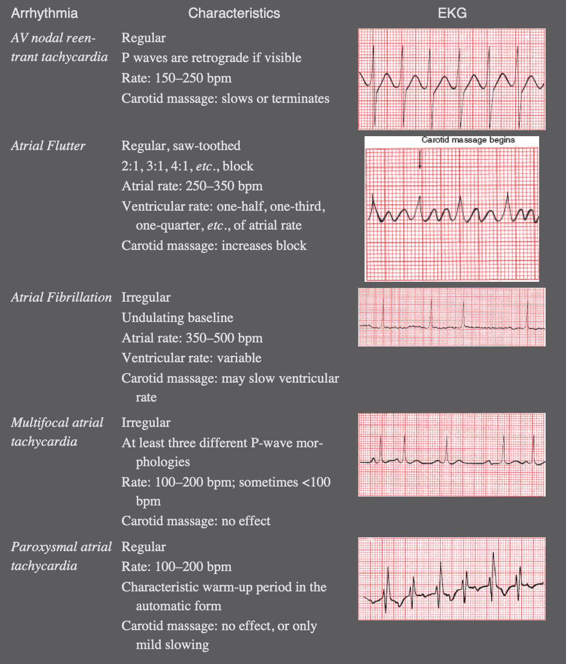
| Regular | AVNRT (PSVT) | P-waves buried/pseudo-R'/retrograde, sudden onset |
|---|
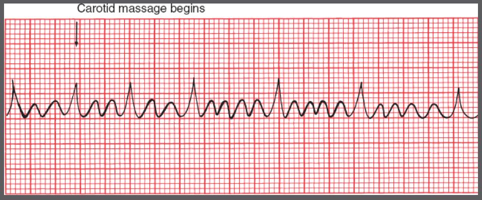
| Regular | Atrial flutter | Saw-tooth flutter waves, 2:1, 3:1, 4:1 conduction |
| Regular | Atrial tachycardia | Abnormal P-waves, may have variable block |
|---|
| Irregularly irregular | Atrial fibrillation | No P-waves, chaotic baseline |
| Irregular | Multifocal atrial tachycardia | ≥3 different P-wave morphologies |
Wide Complex Tachycardia (QRS > 0.12 sec):
- VT vs SVT with aberrancy — This is a critical differentiation
- Clues for VT:
1. AV dissociation (P-waves independent of QRS)
2. Fusion beats
3. Capture beats
4. Concordance (all precordial leads positive or all negative)
5. Extreme axis deviation (-90° to -180°)
6. RS > 100 ms in any precordial lead
7. Positive or negative concordance across precordial leads
- Clues for SVT with aberrancy:
1. P-waves related to QRS (1:1 relationship)
2. Typical RBBB or LBBB pattern
3. Previous EKG with similar aberrancy
Atrial Fibrillation — The 4 Most Common Cardiovascular Causes:
Hypertension
Coronary artery disease (CAD)
Mitral valve disease (especially mitral stenosis)
Heart failure (HF)AVNRT (AV Nodal Reentrant Tachycardia):
- Most common PSVT (regular, narrow-complex, 150-250 bpm)
- Usually triggered by a PAC
- P-waves: Usually buried in QRS (most common), pseudo-R' in V1, or retrograde P-waves in II/III
- Vagal maneuvers (carotid massage) can help diagnose and terminate
- Can occur in healthy hearts with no structural disease
Are normal P-waves present?
Are the QRS complexes narrow or wide?
What is the relationship between P-waves and QRS complexes?
Is the rhythm regular or irregular?Key Distinctions:
- Narrow QRS (≤0.12 sec) → origin at or above AV node
- Wide QRS (>0.12 sec) → origin below AV node (ventricular) or bundle branch block
- Normal P-QRS correlation → rhythm originates in atria (SA node or atrial focus)
- AV dissociation → atria and ventricles are independent
Sinus Rhythms:
| Rhythm | Rate | P-Wave | Regularity |
| -------- | ------ | -------- | ------------ |
|---|
| Normal sinus | 50-100 bpm | Normal, one per QRS | Regular |
| Sinus tachycardia | >100 bpm | Normal | Regular |
|---|
| Sinus bradycardia | <50 bpm | Normal | Regular |
| Sinus arrhythmia | 50-100 bpm | Normal | Slightly irregular (varies with respiration) |
|---|
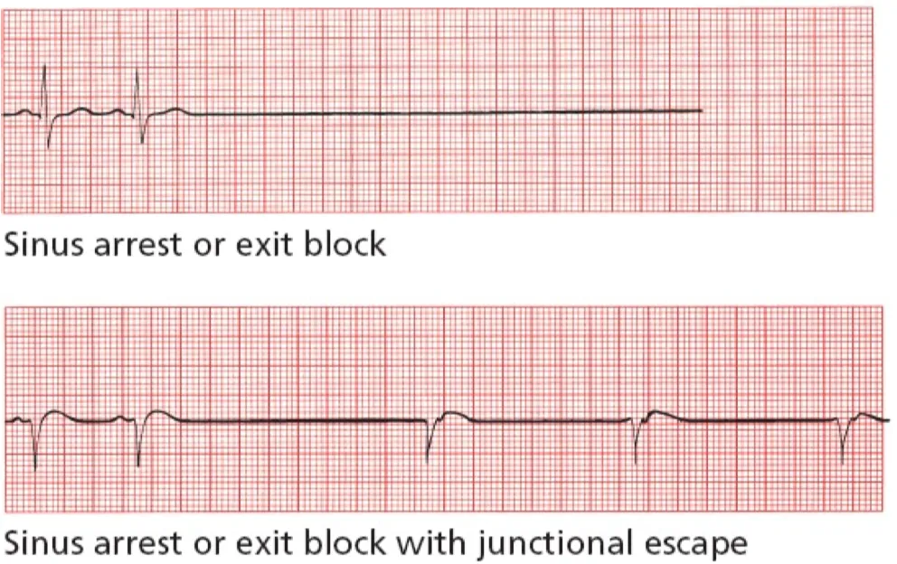
| Sinus arrest/exit block | Variable | Missing pauses | Irregular with pauses |
Atrial Arrhythmias:
| Rhythm | Rate | P-Wave | QRS | Regularity |
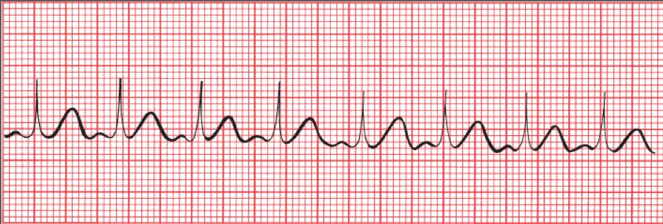
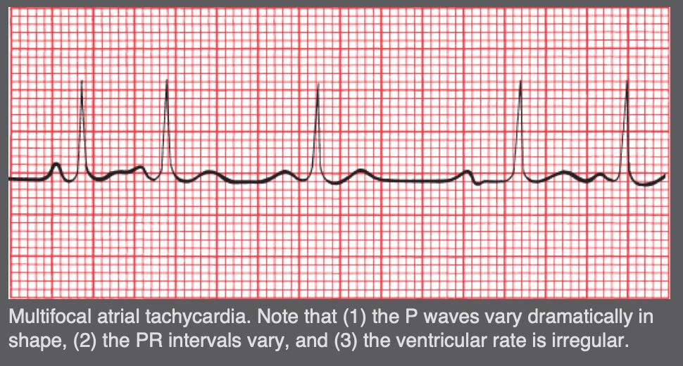
| -------- | ------ | -------- | ----- | ------------ |
|---|
| Atrial fibrillation | Variable | Absent (fibrillatory baseline) | Narrow | Irregularly irregular |
| Atrial flutter | 250-350/min (atrial) | Saw-tooth pattern | Narrow | Usually regular (2:1, 3:1, 4:1 block) |
|---|
| Atrial tachycardia | 150-250/min (atrial) | Abnormal shape, one per QRS | Narrow | Regular |
| AVNRT (PSVT) | 150-250/min | Buried/pseudo-R'/retrograde | Narrow | Regular |
|---|
| WPW/AVRT | Variable | Short PR, delta wave | Narrow or wide | Regular |
| PAC | — | Abnormal P contour | Narrow | Premature, resets rhythm |
AVNRT (AV Nodal Reentry Tachycardia):
- Sudden onset, often triggered by PAC
- Regular, narrow-complex tachycardia (150-250 bpm)
- P-waves buried in QRS (most common), pseudo-R' in V1, or retrograde P-waves in II/III
- Carotid massage can help diagnose and terminate
- Can occur in healthy hearts
Junctional Rhythms:
| Feature | Junctional Escape | Junctional Premature |
|---|
Timing | Late (after pause) | Early (before next expected beat) |
| P-wave | No P-wave or retrograde | No P-wave or retrograde |
|---|
| Rate | 40-60 bpm | — |
| Clinical significance | Benign escape mechanism | Benign |
Premature Beats:
PAC | Atrial | Abnormal P contour | Narrow | Usually resets |
| Junctional premature | Near AV node | Absent/retrograde | Narrow | Usually resets |
|---|
| PVC | Ventricular | None (dissociated) | Wide (>0.12 sec) | Compensatory pause |
Ventricular Arrhythmias:
Ventricular tachycardia (VT) | 100-250 bpm | Wide (>0.12 sec) | Regular |
| Torsades de pointes | 150-250 bpm | Wide, twisting around baseline | Irregular |
|---|
| Ventricular fibrillation | — | Chaotic, no organized QRS | Chaotic |
| PVC | — | Wide, bizarre | Premature with compensatory pause |
|---|
| Ventricular escape | 20-45 bpm | Wide | Late (rescue beats) |
| Accelerated idioventricular rhythm | 45-100 bpm | Wide | Regular |
Heart Block:
| Type | PR-Interval | Dropped Beats | Conduction |
|---|
First-degree AV block | >0.20 sec | None | All conducted, just slow |
| Second-degree Mobitz I (Wenckebach) | Progressive ↑ | Yes | P:QRS ratio increases until dropped |
|---|
| Second-degree Mobitz II | Fixed (normal or prolonged) | Yes | Suddenly dropped without PR lengthening |
| Third-degree (complete) | Variable (dissociated) | Complete | No P-QRS correlation |
|---|
| 2:1 AV block | Variable | Every other P | Can't classify as I or II without longer tracing |
| High-grade AV block | — | ≥2 consecutive dropped | Advanced block |
Atrial Fibrillation:
- Most common and clinically significant sustained arrhythmia
- Key: Irregularly irregular QRS + no discrete P-waves
- Cardiovascular causes: Hypertension, CAD, mitral valve disease, heart failure (PALS-HF mnemonic)
- Lifestyle risk factors: Obesity, excessive alcohol
- Risk of thromboembolism (CHADS₂-VASc score) → anticoagulation
Atrial Flutter:
- Reentrant circuit around tricuspid annulus
- Classic saw-tooth pattern in II, III, aVF
- Counterclockwise (most common): negative flutter waves in II, III, aVF
- Ventricular rate varies with conduction ratio (2:1, 3:1, 4:1)
#### 8. Pre-excitation Syndromes
Wolff-Parkinson-White (WPW):
- Accessory pathway (bundle of Kent) bypassing AV node
- Short PR interval (<0.12 sec) + delta wave (slurred upstroke of QRS) + wide QRS
- Associated tachyarrhythmias: AVRT (most common), atrial fibrillation
- Atrial fibrillation in WPW can degenerate to VF (accessory pathway conducts at very high rates)
- Treatment: Catheter ablation
Lown-Ganong-Levine (LGL):
- Short PR interval WITHOUT delta waves
- Diagnosis requires documented tachyarrhythmia
#### 9. Electrolyte & Drug Effects
Hyperkalemia (Progressive Changes):
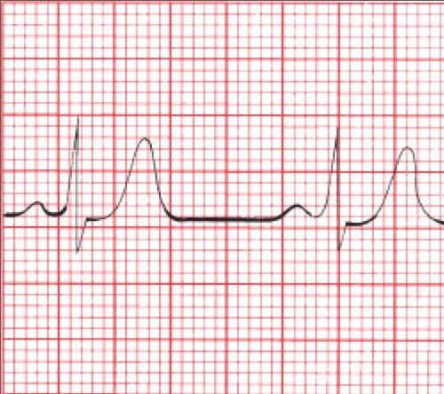
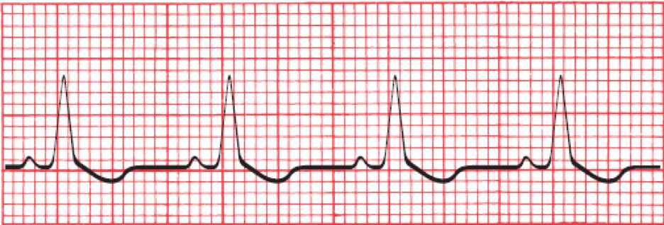
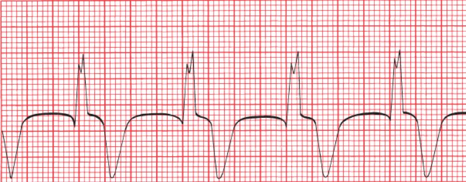
Mild (5.5-6.5) | Tall, peaked T-waves (widespread, all leads) |
| Moderate (6.5-7.5) | PR prolongation → P-wave flattening/disappearance |
|---|
| Severe (7.5-8.5) | Wide QRS, extreme right axis deviation |
| Critical (>8.5) | Sine wave → VF/asystole |
- Hyperkalemia clue: Rightward axis (negative QRS in I, positive in aVF)
Hypokalemia:
Mild (3.0-3.5) | ST depression, flattened T-waves |
| Moderate (2.5-3.0) | U-wave appearance, prolonged QU interval |
|---|
| Severe (<2.5) | ST elevation (rare), arrhythmias |
- U-wave becomes prominent as T-wave flattens
Calcium Abnormalities:
| Abnormality | QT Interval | Risk |
|---|
Hypocalcemia | ↑ Prolonged QT | Torsades de pointes |
| Hypercalcemia | ↓ Shortened QT | — |
Digitalis (Digoxin):
- Therapeutic: ST-segment depression with gradual downslope ("digitalis effect"), T-wave flattening/inversion
- Toxic: PAT (paroxysmal atrial tachycardia) with block is most characteristic; also PVCs, junctional tachy, AV block
- Slows conduction through AV node
- Can suppress sinus node
#### 10. Pericarditis & Other Conditions
Acute Pericarditis:
- Diffuse (widespread) ST-segment elevation, concave upward
- PR-interval depression (early sign)
- Later T-wave flattening/inversion
- No reciprocal changes (unlike STEMI)
- Differentiate from STEMI: Pericarditis is diffuse (not in a single territory), concave upward
Pericardial Effusion:
- Low voltage in all leads (QRS <0.5 mV limb, <1.0 mV precordial)
- Electrical alternans (alternating QRS amplitude) — highly specific
Pulmonary Embolism:
- S1Q3T3 pattern (S in I, Q in III, T inversion in III)
- RVH pattern, right axis deviation
- Right bundle branch block (may be transient)
- Sinus tachycardia
Chronic Obstructive Pulmonary Disease (COPD):
- Low voltage
- Right axis deviation
- Poor R-wave progression in precordial leads
Hypothermia:
- Bradycardia
- Prolongation of all intervals
- Osborn waves (J waves) — prominent deflection at J point, best seen in II, V5-V6
- Muscle tremor artifact (may simulate atrial flutter/flutter)
Brugada Pattern:
V1-V3 | ST elevation
 (coved type) |
| V1 | RBBB pattern with slow descent of R' |
|---|
| V1-V2 | T-wave inversion |
| Clinical | Risk of sudden cardiac death (Brugada syndrome) |
Central Nervous System (CNS) Events:
- Subarachnoid bleed, CVA, head trauma
- Diffuse T-wave inversions plus QT prolongation
- Prominent U-waves
#### 11. Systematic 9-Step EKG Interpretation
Heart rate: Calculate (300 method or 6-second method)
Rhythm: Regular? P-waves present? P-QRS relationship?
Axis: Check I and aVF → normal, LAD, or RAD
Intervals: PR, QRS, QT
Conduction blocks: LBBB, RBBB, fascicular, AV block
Hypertrophy/Enlargement: LVH, RVH, atrial enlargement
ST-T Changes: Ischemia, injury, electrolyte, digoxin, pericarditis
Q-waves: Significant? Pathological?
Comparison with prior EKG: New changes?